Learning Objectives
Master the anatomy and pathophysiology of Rhinosinusitis. Understand the drainage pathways of the paranasal sinuses, the common microbial triggers, and the critical anatomical complications involving the orbits and cavernous sinus for the USMLE Step 1.
1. Pathophysiology and Sinus Anatomy
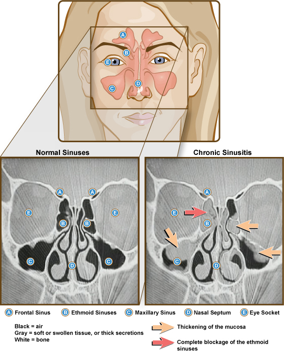
Rhinosinusitis involves the obstruction of sinus drainage into the nasal cavity, leading to inflammation and localized pain. The Maxillary sinus is the most commonly affected due to its unique drainage mechanics.
| Sinus | Drainage Pathway | Clinical Note |
|---|---|---|
| Maxillary | Middle Meatus (via superomedial ostia) | Drains against gravity; high risk of fluid stasis. |
| Frontal / Ant. Ethmoid | Middle Meatus | Pain is often felt in the forehead or between the eyes. |
| Posterior Ethmoid | Superior Meatus | Deep facial pain or pressure. |
| Nasolacrimal Duct | Inferior Meatus | Blockage leads to epiphora (excessive tearing). |
2. Microbiology of Acute Rhinosinusitis
Most cases of acute rhinosinusitis are viral, but secondary bacterial infections are a common high-yield topic.
| Type | Common Pathogens |
|---|---|
| Viral (Most Common) | Rhinovirus, Influenza, Parainfluenza. |
| Bacterial (Superimposed) | 1. S. pneumoniae
2. Nontypeable H. influenzae 3. M. catarrhalis |
3. Anatomical Complications
Because the paranasal sinuses are in proximity to vital structures, infections can spread rapidly if not managed.
| Complication | Anatomical Extension | Clinical Signs |
|---|---|---|
| Orbital Cellulitis | Through the thin lamina papyracea of the ethmoid bone. | Proptosis, painful eye movement, and vision loss. |
| Cavernous Sinus Syndrome | Posterior extension via venous drainage. | Cranial nerve palsies (III, IV, V1, V2, VI). |
| Meningitis/Brain Abscess | Superior extension through the cribriform plate. | Fever, headache, nuchal rigidity, focal neuro deficits. |
Activity:
High-Yield Clinical Pearls:
- The Middle Meatus: Remember that most major sinuses (frontal, maxillary, anterior ethmoid) drain here. It is the “hub” of sinus pathology.
- Gravity Challenge: Maxillary sinusitis is unique because the ostium is located at the top of the sinus. You basically have to fill the “bucket” before it can drain.
- Fungal Alert: In immunocompromised patients (e.g., DKA, leukemia), always consider Mucor or Rhizopus as a cause of necrotizing rhinosinusitis.
You must be logged in to post a comment.