
Learning Objectives
- Detail the developmental anatomy and structural hallmarks of major posterior fossa malformations, including Chiari I, Chiari II, and Dandy–Walker anomalies.
- Differentiate between adult and infant clinical presentations, recognizing symptoms of brainstem compression, cerebellar ataxia, and elevated intracranial pressure.
- Correlate specific posterior fossa anomalies with associated congenital pathologies such as syringomyelia and myelomeningocele.
- Evaluate imaging findings and the development of obstructive (non-communicating) hydrocephalus resulting from cerebrospinal fluid pathway blockage.
1. Summary of Posterior Fossa Malformations
Congenital structural defects of the cerebellum and brainstem produce distinct clinical syndromes depending on the degree of herniation or dysgenesis.
| Malformation | Anatomical Defects | Clinical Presentation | Associated Conditions |
|---|---|---|---|
| Chiari I Malformation | Caudal displacement of the cerebellar tonsils through the foramen magnum. | Often asymptomatic in childhood; manifests in adulthood with occipital headaches, neck pain, and cerebellar ataxia. | Syringomyelia (central spinal cord cavitation) |
| Chiari II Malformation
A |
Downward displacement of the cerebellar vermis, tonsils, and medulla through the foramen magnum. | Infant presentation: dysphagia, stridor, apnea, and limb weakness due to severe brainstem compression. | Myelomeningocele (lumbosacral spina bifida) & obstructive hydrocephalus |
| Dandy–Walker Malformation
B |
Agenesis of the cerebellar vermis combined with massive cystic dilation of the 4th ventricle. | Delayed motor development, hypotonia, and signs of increased intracranial pressure. | Noncommunicating hydrocephalus & spina bifida |
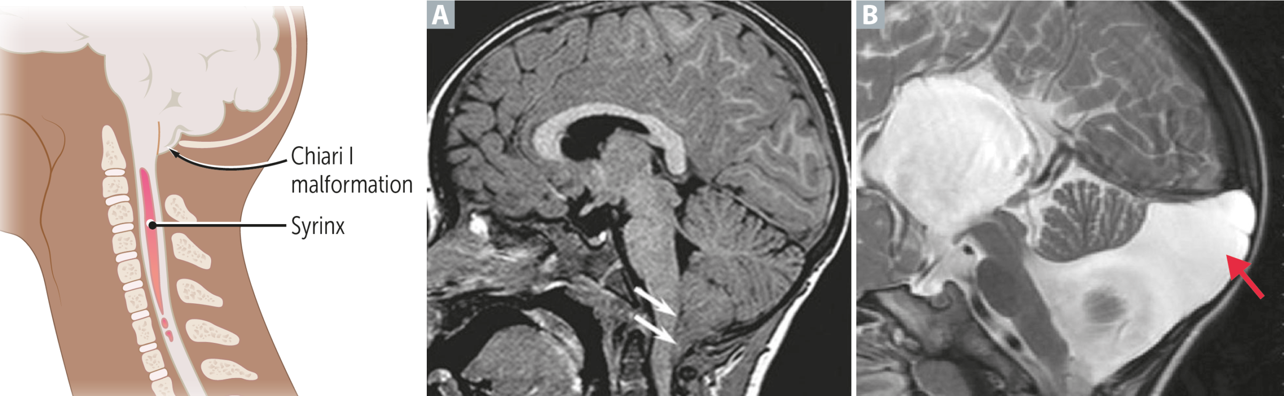
Clinical Notes & Pathology:
- Chiari I & Syringomyelia: Tonsillar herniation obstructs CSF outflow from the fourth ventricle into the subarachnoid space, driving fluid into the central spinal canal and forming a syrinx that causes “cape-like” loss of pain and temperature sensation.
- Chiari II Severity: The inclusion of brainstem structures (medulla) makes Chiari II significantly more dangerous, frequently requiring surgical decompression in infancy.
- Dandy–Walker Dynamics: The expansion of the fourth ventricle enlarges the posterior fossa, preventing normal cerebellar vermis development and disrupting CSF resorption.
Key Points & Memory Hooks:
Chiari I vs. II: Think “tonsils alone” for Chiari I (presents later in adults with syringomyelia) versus “vermis + tonsils + medulla” for Chiari II (presents early in infants with myelomeningocele).
Dandy–Walker Hallmark: Absent vermis + giant cystic fourth ventricle.
Imaging Choice: MRI is the gold standard modality for visualizing soft tissue herniation and spinal cord abnormalities.
Chiari I vs. II: Think “tonsils alone” for Chiari I (presents later in adults with syringomyelia) versus “vermis + tonsils + medulla” for Chiari II (presents early in infants with myelomeningocele).
Dandy–Walker Hallmark: Absent vermis + giant cystic fourth ventricle.
Imaging Choice: MRI is the gold standard modality for visualizing soft tissue herniation and spinal cord abnormalities.
You must be logged in to post a comment.