
The Molecular Basis of Cancer: Regulatory Pathways
At its core, cancer is a genetic disease characterized by a progressive accumulation of non-lethal mutations. These genetic alterations selectively target regulatory pathways that dictate cellular life, division, and death, shifting a normal cell line into an autonomous, malignant clone.
1. The Genetic Tug-of-War: Oncogenes vs. Tumor Suppressors
Malignant transformation requires a dual disruption: activating the genetic accelerators of cell division while simultaneously dismantling the biochemical brakes.
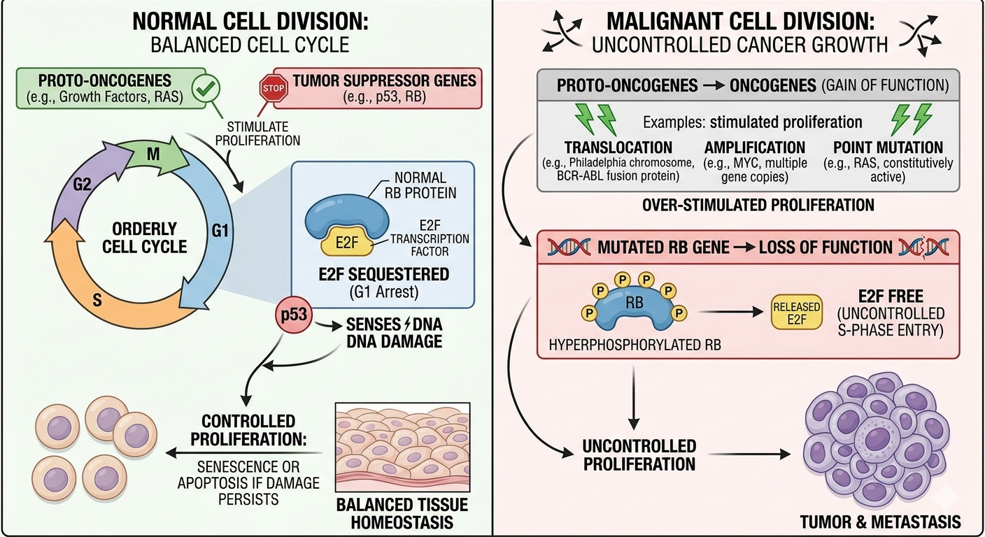
| Gene Class | Molecular Mechanism | High-Yield Clinical Examples & Associations |
|---|---|---|
| RAS | GTPase signaling protein. Point mutations lock RAS in its active, GTP-bound state, sending continuous proliferation signals to the nucleus. | The most common oncogene mutation in human tumors. Associated with Pancreatic Adenocarcinoma and Colorectal Cancer. |
| MYC | A transcription factor that upregulates pro-growth cyclins and metabolic enzymes. | • c-MYC: Translocation t(8;14) in Burkitt Lymphoma. • n-MYC: Amplification in Neuroblastoma. |
| TP53 | Guardian of the Genome. Activated by DNA damage to induce cell cycle arrest via p21 (G1/S checkpoint) or trigger apoptosis via BAX/BAK. | Germline mutations cause Li-Fraumeni Syndrome (multiple early-onset primary sarcomas, breast, brain, and adrenal carcinomas). |
| Rb | Governor of the Cell Cycle. Hypophosphorylated Rb binds and sequesters E2F transcription factors, blocking entry into S-phase. | Inactivated by hyperphosphorylation or viral proteins (HPV E7). Germline mutations drive familial Retinoblastoma and Osteosarcoma. |
2. Cell Cycle Regulation and the G1/S Checkpoint
Progression through the cell cycle is tightly orchestrated by Cyclins and Cyclin-Dependent Kinases (CDKs). The decision to commit to DNA replication occurs exclusively at the G1/S checkpoint:
- The Cyclin D/CDK4 Complex: Growth factor pathways drive the accumulation of Cyclin D, which binds and activates CDK4 and CDK6. This active complex phosphorylates the Rb protein.
- E2F Liberation: Once Rb is hyperphosphorylated, it undergoes a conformational shift and releases E2F. Free E2F translates genes required for the S-phase, including Cyclin E and DNA polymerase.
- CDK Inhibitors (CDKIs): Tumor suppressors regulate this checkpoint by utilizing proteins like p16, p21, and p27 to lock CDKs in an inactive state, preventing premature Rb phosphorylation.
3. DNA Repair Pathways & Genomic Instability
When cells lose their capacity to patch damaged DNA, the pace of carcinogenesis accelerates exponentially. These defects fall into three primary repair systems:
- Mismatch Repair (MMR): Fixes single-base mismatches and small insertion/deletion loops that slip past DNA polymerase during replication.
• Defect: Loss of MMR genes (MLH1, MSH2, MSH6, PMS2) causes **Microsatellite Instability (MSI)** and drives Lynch Syndrome (Hereditary Non-Polyposis Colorectal Cancer).
- Nucleotide Excision Repair (NER): Excises bulky, helix-distorting lesions, most notably pyrimidine dimers formed by ultraviolet (UV) radiation.
• Defect: Inactivating mutations in NER pathways lead to Xeroderma Pigmentosum, predisposing patients to severe skin carcinomas under minimal sun exposure.
- Homologous Recombination: Resolves lethal double-strand DNA breaks using the sister chromatid as an error-free template.
• Defect: Inactivation of BRCA1 or BRCA2 selectively cripples this pathway, massively increasing life-time risks for hereditary breast, ovarian, and prostatic adenocarcinomas.
You must be logged in to post a comment.