
Common Cancer-Associated Mutations: Oncogenes, Suppressors, & Translocations
Malignant transformations are driven by specific genetic alterations that can be categorized into gain-of-function oncogenic activations, loss-of-function tumor suppressor inactivation, and recurrent chromosomal translocations. Recognizing these specific genetic markers is crucial for both diagnosis and targeted therapeutic selection.
1. High-Yield Proto-Oncogenes (Gain of Function)
Proto-oncogenes require a mutation in only one allele to become constitutively active, sending continuous growth and survival signals to the cell nucleus.
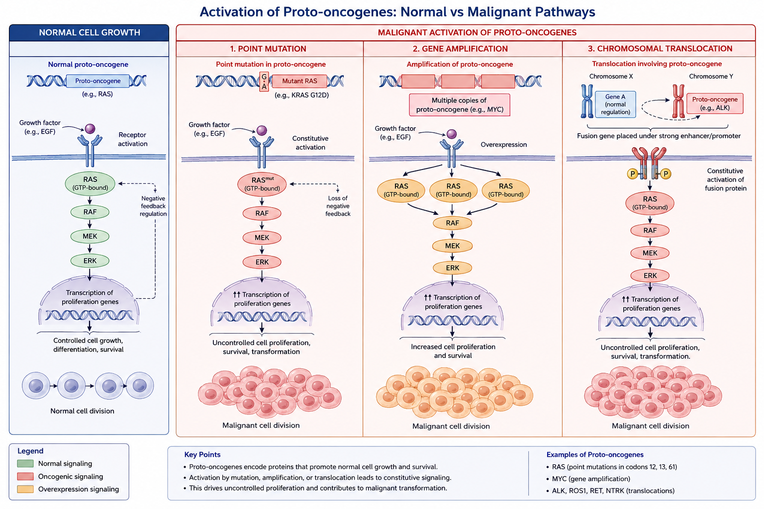
| Oncogene | Mutation Mechanism & Function | Associated Malignancies & Targeted Therapy |
|---|---|---|
| ALK | Receptor tyrosine kinase inversion mutation on chromosome 2, forming the EML4-ALK fusion protein. | Seen in Non-Small Cell Lung Cancer (NSCLC) in non-smokers. Targeted with Crizotinib or Alectinib. |
| BRAF | Serine/threonine kinase point mutation (V600E: valine to glutamic acid replacement). | Characteristically seen in Melanoma, Papillary Thyroid Carcinoma, and Hairy Cell Leukemia. Targeted with Vemurafenib. |
| EGFR | Receptor tyrosine kinase deletions or point mutations driving autonomous activation. | Common in lung adenocarcinoma (especially in Asian females who are non-smokers). Targeted with Erlotinib or Osimertinib. |
| HER2 (ERBB2) | Receptor tyrosine kinase gene amplification leading to massive protein overexpression. | Associated with aggressive subtypes of Breast Carcinoma and gastric carcinomas. Targeted with Trastuzumab. |
| KRAS / NRAS | GTPase point mutations that block GTP hydrolysis, locking RAS in a permanent active signaling state. | Prevalent in colorectal, pancreatic, and lung cancers. The presence of KRAS mutations predicts resistance to anti-EGFR therapies like Cetuximab. |
| RET | Receptor tyrosine kinase point mutations or fusions. | Germline mutations drive Multiple Endocrine Neoplasia type 2A and 2B (MEN 2A/2B) and Medullary Thyroid Carcinoma. |
2. High-Yield Tumor Suppressor Genes (Loss of Function)
Tumor suppressors typically comply with Knudson’s “two-hit hypothesis,” meaning functional inactivation requires a loss-of-function mutation in both cellular alleles.
- APC (Adenomatous Polyposis Coli): Regulates cell adhesion and downregulates beta-catenin within the Wnt signaling pathway. Loss of APC leads to unchecked beta-catenin accumulation, driving transcription of growth-promoting genes. Germline mutations result in Familial Adenomatous Polyposis (FAP), where thousands of colonic polyps develop by early adulthood.
- BRCA1 / BRCA2: Code for proteins essential to the homologous recombination pathway for double-stranded DNA break repair. Inactivation leads to severe genomic instability. Inherited mutations dramatically elevate the lifetime incidence of early-onset Breast and Ovarian Carcinomas in women and pancreatic or prostate cancers in men.
- VHL (Von Hippel-Lindau): Part of a ubiquitin ligase complex that targets Hypoxia-Inducible Factor 1-alpha (HIF-1a) for proteasomal destruction under normoxic conditions. Loss of VHL prevents HIF-1a degradation, mimicking chronic hypoxia and driving runaway transcription of VEGF and erythropoietin. Associated with Clear Cell Renal Cell Carcinoma, hemangioblastomas, and pheochromocytomas.
- WT1 (Wilms Tumor 1): A zinc-finger transcription factor vital for normal urogenital development. Inactivating mutations or deletions on chromosome 11p13 lead to the development of Wilms Tumor (Nephroblastoma) in pediatric patients, either isolated or as part of WAGR syndrome (Wilms tumor, Aniridia, Genitourinary anomalies, and intellectual disability).
3. Recurrent Chromosomal Translocations to Memorize
Specific recurrent structural rearrangements are pathognomonic for specific hematologic and soft-tissue malignancies:
| Translocation | Fusion Gene / Alteration | Malignancy & Clinical Correlation |
|---|---|---|
| t(9;22) | BCR-ABL1 fusion (Philadelphia chromosome) | Creates a constitutively active tyrosine kinase. Diagnostic for Chronic Myelogenous Leukemia (CML); also found in a poor-prognosis subset of Acute Lymphoblastic Leukemia (ALL). Treated with Imatinib. |
| t(15;17) | PML-RARA fusion | Disrupts the retinoic acid receptor, blocking promyelocytic differentiation. Diagnostic for Acute Promyelocytic Leukemia (APL, AML M3). High risk of DIC. Treated with **All-Trans Retinoic Acid (ATRA)**. |
| t(8;14) | c-MYC moved to the heavy-chain Ig locus | Places the c-MYC transcription factor under the control of a highly active immunoglobulin promoter, driving rapid cell division. Diagnostic for Burkitt Lymphoma. |
| t(14;18) | BCL-2 moved to the heavy-chain Ig locus | Upregulates the anti-apoptotic protein BCL-2, preventing normal programmed cell death in B-cells. Diagnostic for Follicular Lymphoma. |
| t(11;14) | Cyclin D1 moved to the heavy-chain Ig locus | Upregulates Cyclin D1, driving rapid hyperphosphorylation of Rb and unregulated passage through the G1/S cell cycle checkpoint. Diagnostic for Mantle Cell Lymphoma. |
| t(11;22) | EWSR1-FLI1 fusion | Creates an oncogenic transcription factor. Diagnostic for Ewing Sarcoma (malignant round-blue-cell bone tumor in pediatric populations). |
You must be logged in to post a comment.