
Learning Objectives
- Describe the anatomical location, structure, and cellular composition (pinealocytes and glial cells) of the pineal gland.
- Explain the synthesis and circadian regulation of melatonin and its role in sleep-wake cycles.
- Identify clinical conditions associated with the gland, including circadian rhythm disorders, pineal tumors, pineal cysts, and age-related calcification.
1. Anatomy & Cellular Structure
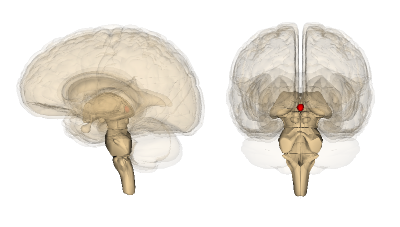
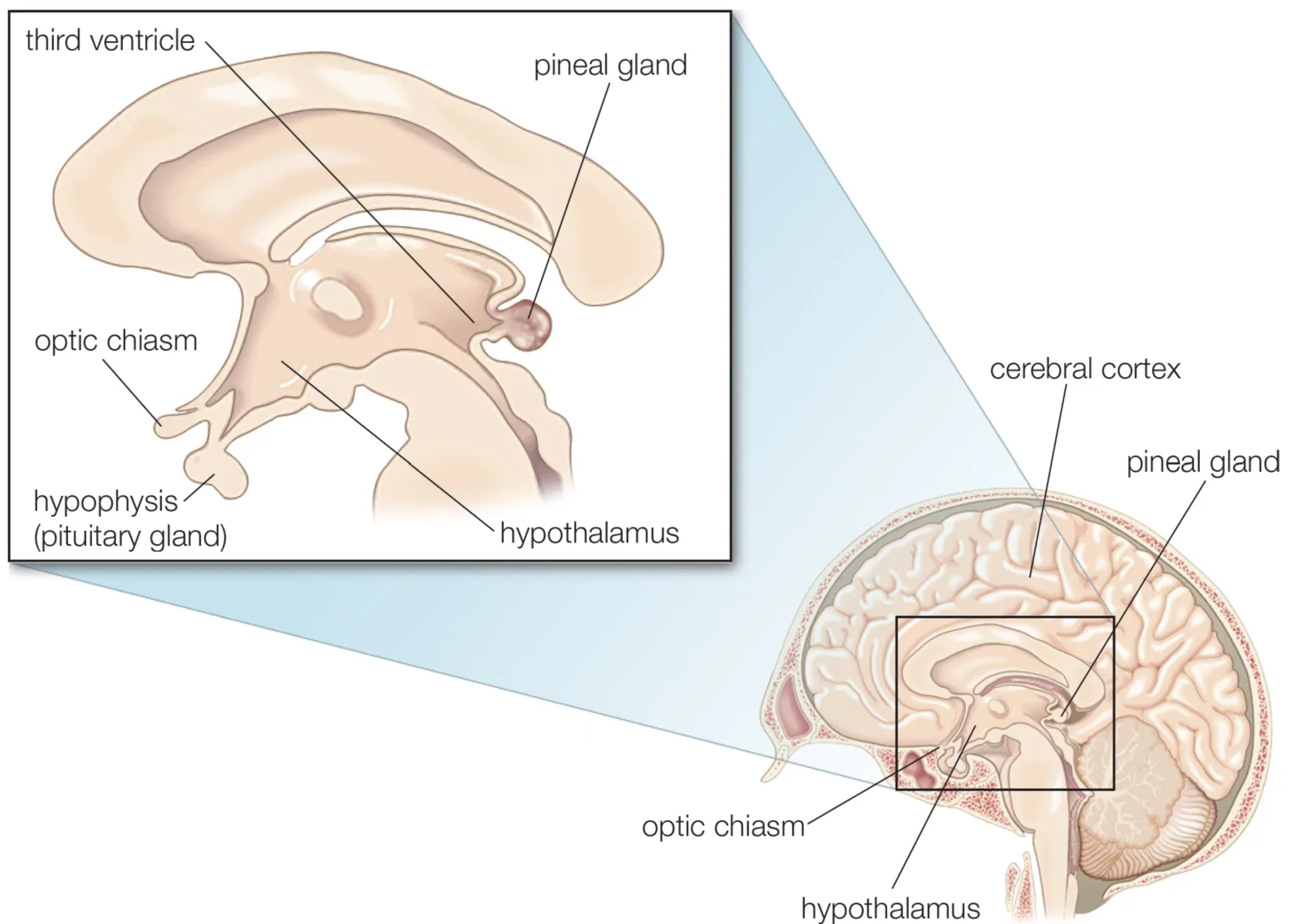
The pineal gland (pineal body or epiphysis) is a small, pinecone-shaped endocrine gland measuring approximately 8 mm in length, located near the brain’s centerline.
- Location: Situated in the epithalamus between the two cerebral hemispheres, just above the brainstem.
- Cellular Composition: Composed of pinealocytes (the primary functional hormone-producing cells) supported by neuroglial cells.
2. Hormonal Regulation & Melatonin
- Melatonin Production: The gland’s primary function is the synthesis and secretion of melatonin, governed by light-dark cycles via the circadian rhythm.
- Light Inhibition: Melatonin levels peak during darkness to promote sleep and adjust seasonal responses, while light exposure suppresses its production.
Clinical Notes & Pathology:
- Circadian Disorders: Melatonin dysregulation contributes to insomnia, jet lag, delayed sleep phase syndrome, and Seasonal Affective Disorder (SAD).
- Pineal Tumors & Cysts: Neoplasms (benign pineocytomas or malignant pineoblastomas) and large fluid-filled cysts can compress surrounding midbrain structures (such as the tectal plate), leading to headaches, vision problems, and hydrocephalus.
- Calcification: Age-related calcium deposition within the pineal gland is a common, typically benign incidental finding on neuroimaging.
Key Points & Memory Hooks:
Darkness Hormone: Melatonin is your biological night signal, synthesized by pinealocytes when light input drops.
Epithalamus Anchor: The pineal body sits centrally as the anatomical landmark of the epithalamus.
Mass Effect Warning: Because of its tight quarters near the cerebral aqueduct, pineal masses frequently obstruct CSF flow to cause hydrocephalus.
Darkness Hormone: Melatonin is your biological night signal, synthesized by pinealocytes when light input drops.
Epithalamus Anchor: The pineal body sits centrally as the anatomical landmark of the epithalamus.
Mass Effect Warning: Because of its tight quarters near the cerebral aqueduct, pineal masses frequently obstruct CSF flow to cause hydrocephalus.
You must be logged in to post a comment.