
Learning Objectives
- Understand the structure, bony architecture, and functional divisions of the palate.
- Identify the specific soft palate muscles, their neural innervation, and their precise roles during swallowing and vocalization.
- Analyze the neurovascular supply, foraminal landmarks, and clinical relevance of developmental defects such as cleft lip and cleft palate.
Anatomical Position & Hard Palate Architecture
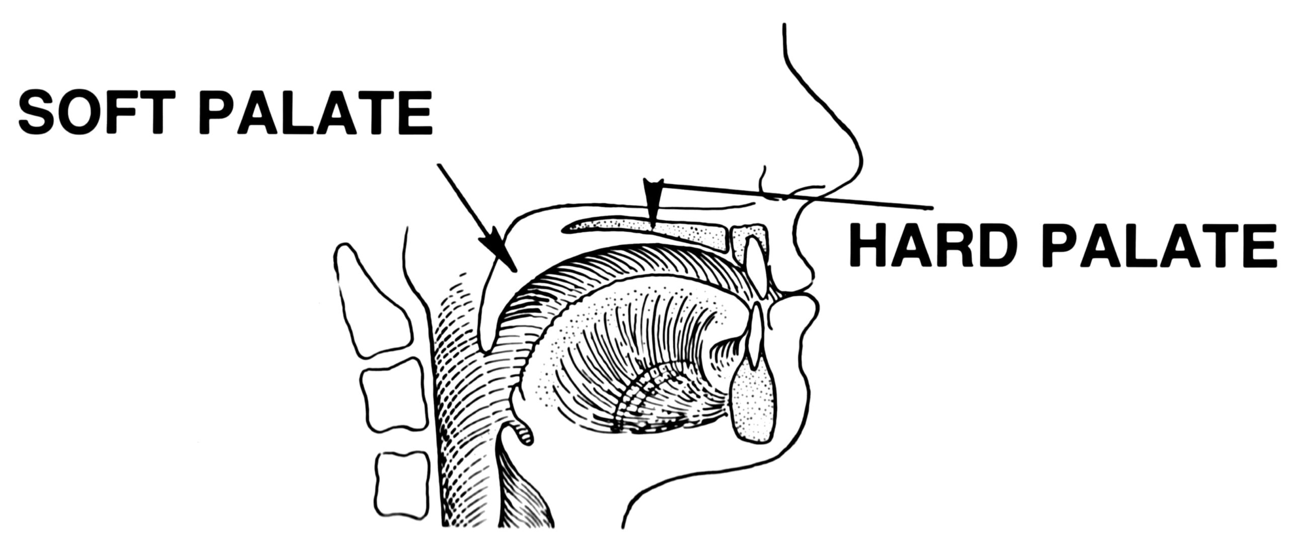
The palate forms the roof of the mouth and separates the nasal cavity from the oral cavity. Its superior surface is covered by respiratory epithelium, while the inferior surface features oral mucosa containing salivary glands. The hard palate is a rigid, bony structure formed by distinct skeletal components:
| Palatal Component | Bony Framework & Foramina | Neurovascular & Clinical Significance |
|---|---|---|
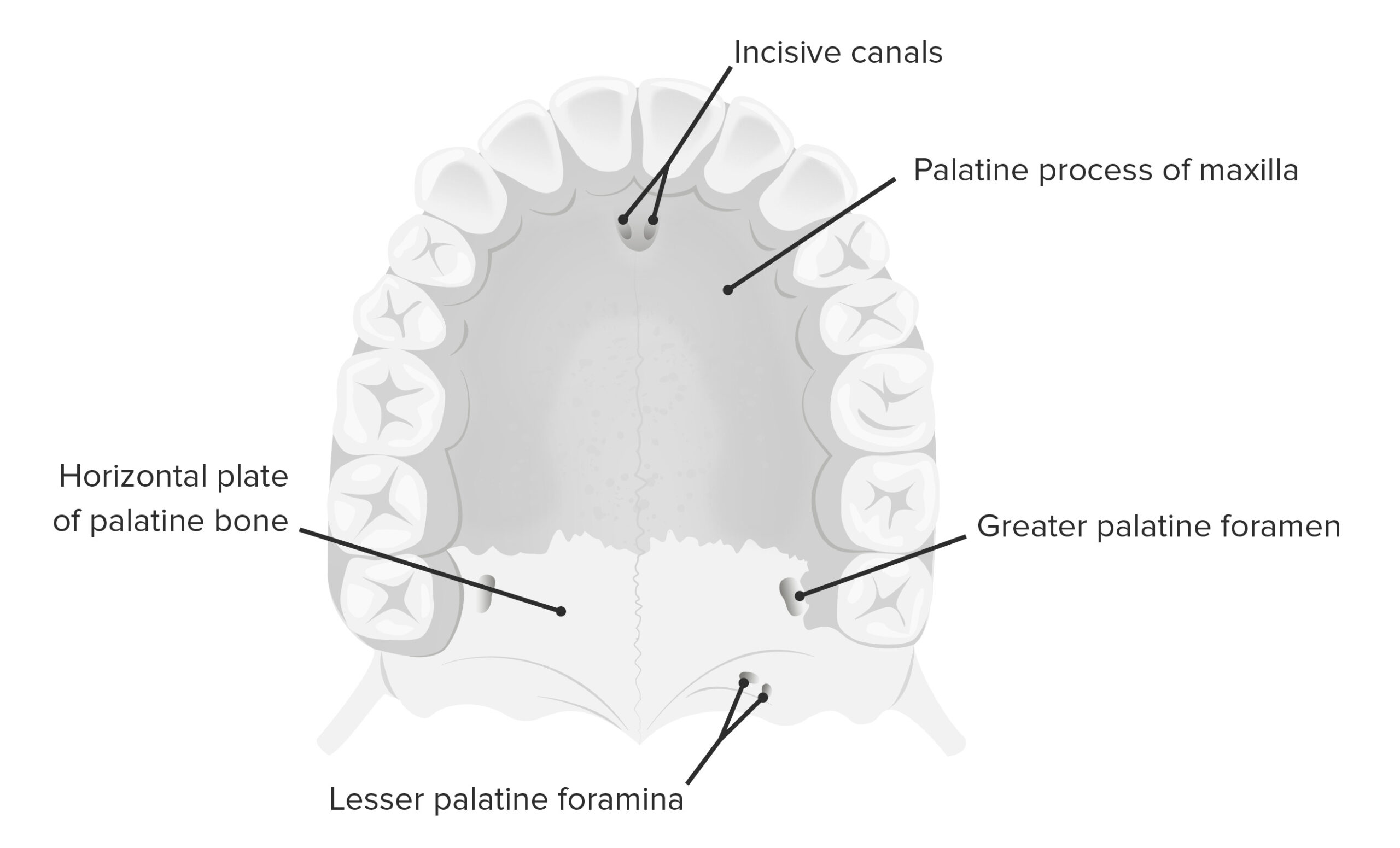
| Hard Palate Bones | Palatine processes of the maxilla & horizontal plates of the palatine bones | Provides a stable, immobile bony partition between the oral and nasal cavities. |
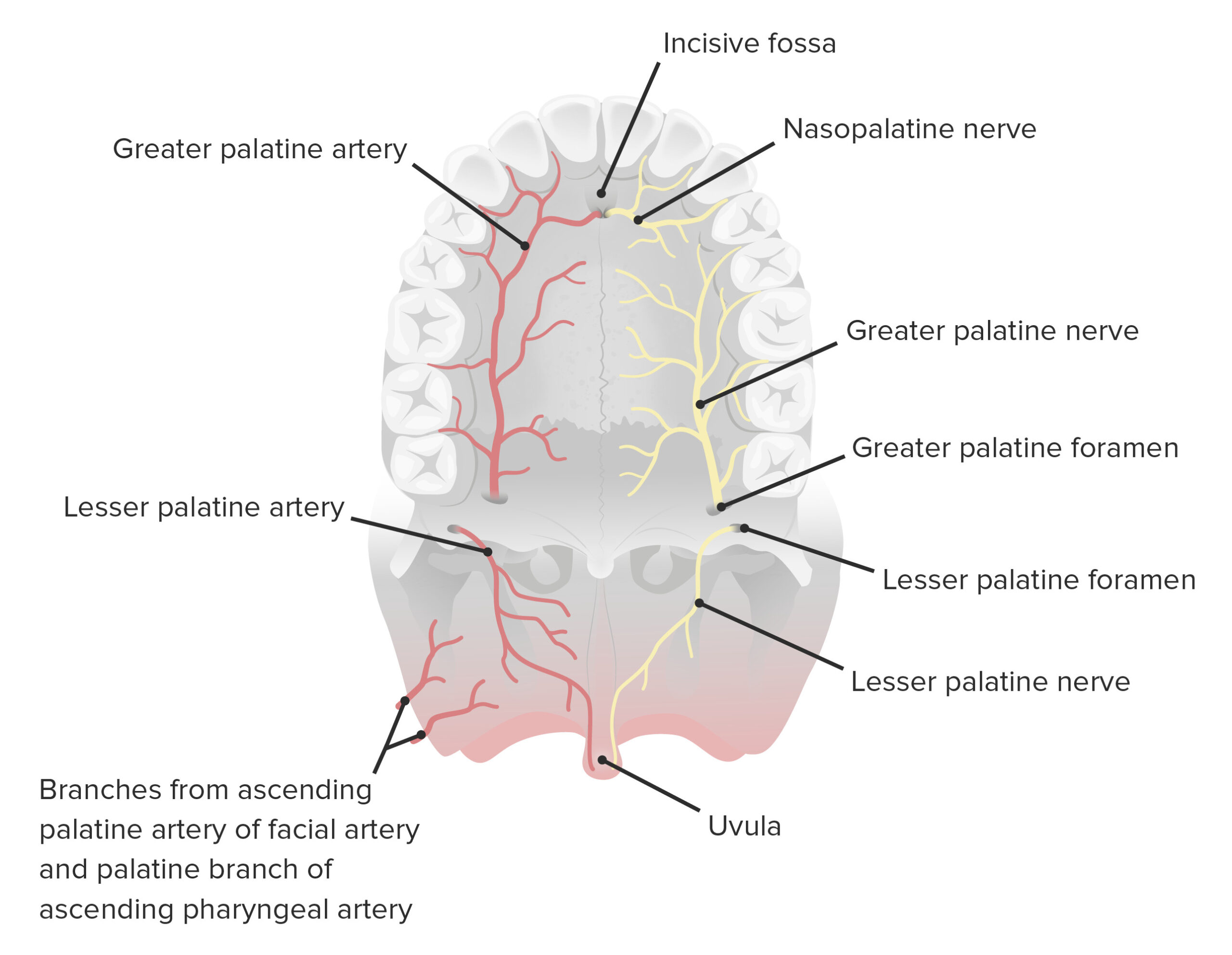
| Incisive Canal | Anterior bony aperture in the hard palate | Transmits the nasopalatine nerve and descending palatine artery. |
| Greater/Lesser Palatine Foramina | Posterolateral openings in the palatine bone | Transmits the greater/lesser palatine nerves and accompanying vessels. |
Soft Palate Muscles & Innervation
The soft palate is a mobile, muscular structure continuous with the palatine aponeurosis, terminating in the uvula and forming the roof of the fauces (bounded by the palatoglossal and palatopharyngeal arches, enclosing the palatine tonsils):
| Muscle Name | Primary Functional Action | Neural Innervation |
|---|---|---|
| Tensor Veli Palatini | Tenses the soft palate | Mandibular nerve ( ) |
| Levator Veli Palatini | Elevates the soft palate during swallowing | Vagus nerve ( ) |
| Palatoglossus | Pulls the palate toward the tongue | Vagus nerve () |
| Palatopharyngeus | Tenses palate; draws pharynx anteriorly | Vagus nerve () |
| Musculus Uvulae | Shortens the uvula | Vagus nerve () |
Clinical Relevance – Cleft Lip, Cleft Palate & Vascular Supply
Vascular Pathways & Developmental Anomalies
- Arterial Supply & Drainage: Supplied primarily by the greater palatine artery, with anastomoses from the lesser palatine and ascending palatine arteries; venous drainage flows into the pterygoid venous plexus.
- Cleft Lip: Developmental defect resulting from failed fusion of the medial nasal prominence and the maxillary prominence.
- Cleft Palate: Caused by failed fusion of the palatal shelves; occurs independently or alongside cleft lip, leading to feeding difficulties, speech issues, and recurrent infections.
- Epidemiology: Occurs in approximately 1 in 1,000 live births, with a significantly higher incidence in specific populations such as Native Americans.
Key Points & Memory Hooks:
- Core Rule: Almost all soft palate muscles are innervated by the vagus nerve (
- Memory Hook: Tensor is a Trigeminal outlier—
tenses the palate before the vagus takes over the rest!
You must be logged in to post a comment.