
Learning Objectives
- Describe the five layers of the scalp using the SCALP mnemonic.
- Identify the arterial supply and venous drainage of the scalp.
- Distinguish cutaneous innervation from the trigeminal nerve and cervical nerves.
- Recognize the clinical relevance of the loose areolar tissue as the danger area.
1. Layers of the Scalp
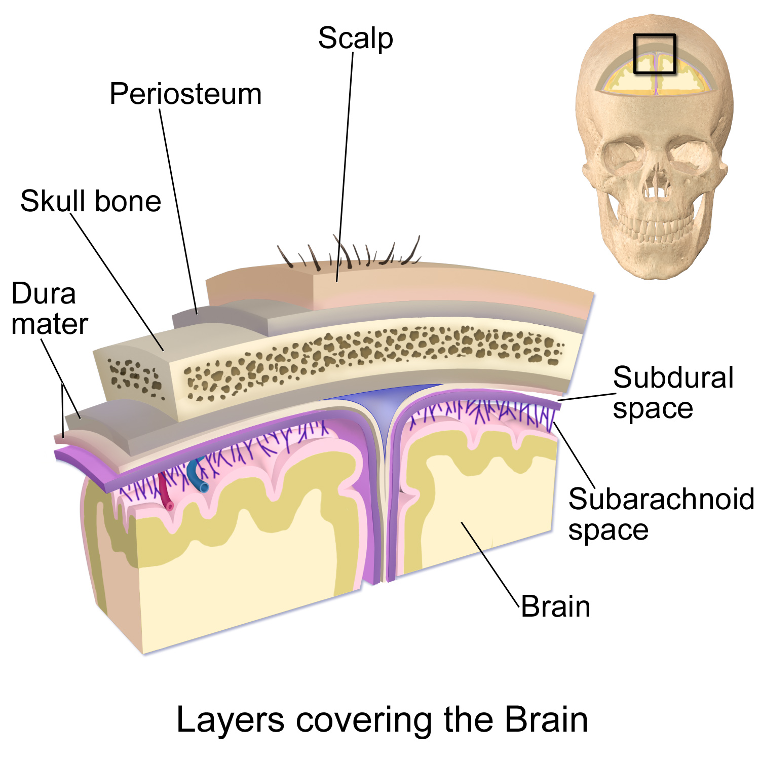
The scalp refers to the layers of skin and subcutaneous tissue covering the cranial vault, serving protective, sensory, and thermoregulatory functions. It consists of five layers, remembered by the mnemonic SCALP:
- S – Skin: Contains hair follicles and sebaceous glands, making it a common site for sebaceous cysts.
- C – Dense Connective Tissue: Connects skin to the epicranial aponeurosis. Richly vascularized and innervated; tightly bound vessels prevent effective vasoconstriction, leading to profuse bleeding in wounds.
- A – Aponeurosis (Epicranial): A thin, tendon-like structure connecting the occipitalis and frontalis muscles, allowing coordinated movement of the scalp.
- L – Loose Areolar Connective Tissue: Known as the danger area of the scalp; contains emissary veins connecting superficial veins to intracranial venous sinuses.
- P – Periosteum: The outer covering of cranial bones; continuous with the endosteum at sutural margins.
2. Neurovascular Supply
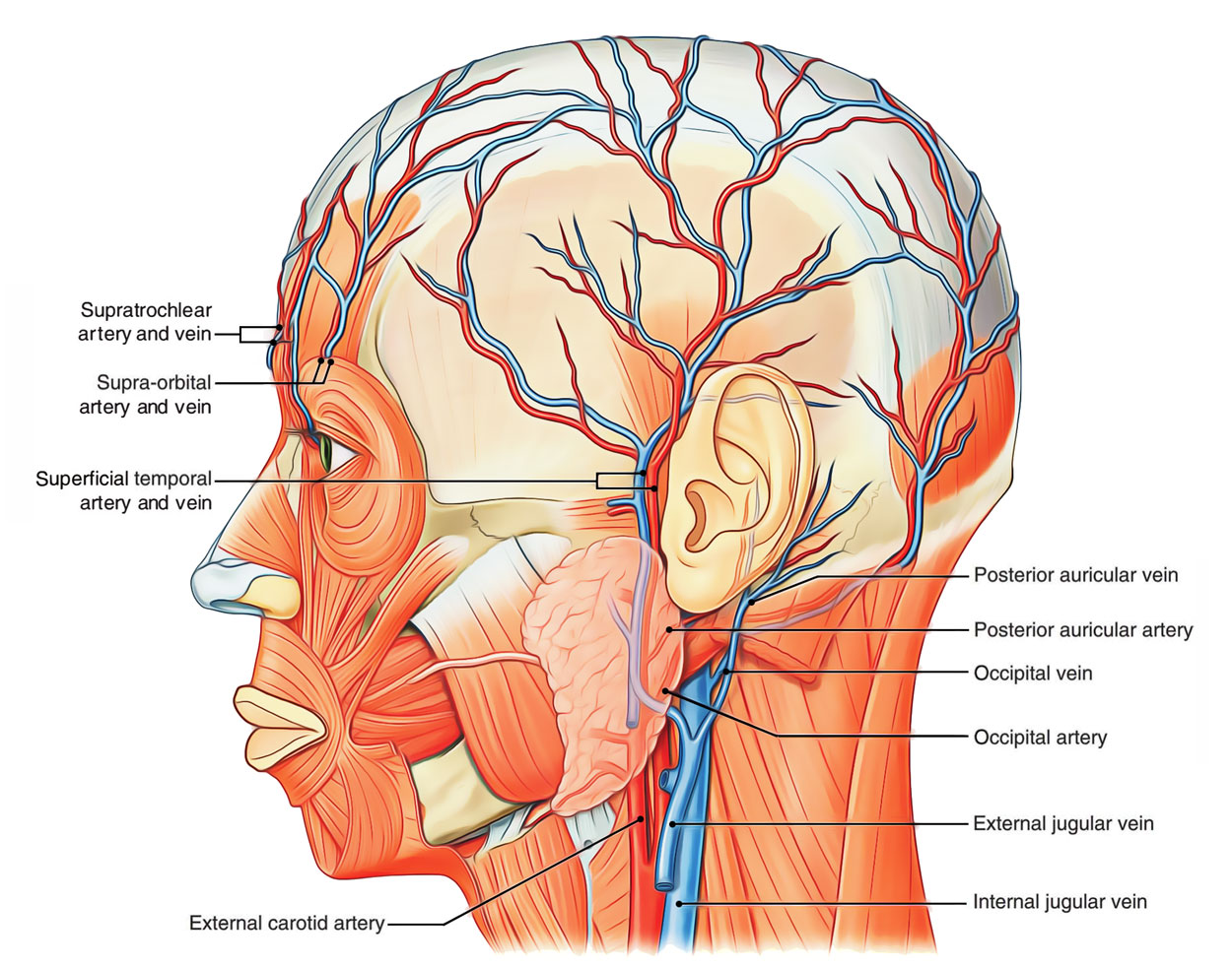
The scalp receives extensive blood supply from both external and internal carotid arteries:
- External Carotid Branches:
- Superficial temporal artery – frontal and temporal regions
- Posterior auricular artery – area above and behind the ear
- Occipital artery – posterior scalp
- Internal Carotid Branches (via Ophthalmic artery):
- Supraorbital artery
- Supratrochlear artery
Venous Drainage:
- Superficial veins: Follow the arterial pattern (superficial temporal, occipital, posterior auricular, supraorbital, supratrochlear).
- Deep drainage: Via the pterygoid venous plexus, connecting to the maxillary vein.
- Emissary veins: Connect to diploic and dural venous sinuses, providing routes for infection spread.
3. Innervation of the Scalp
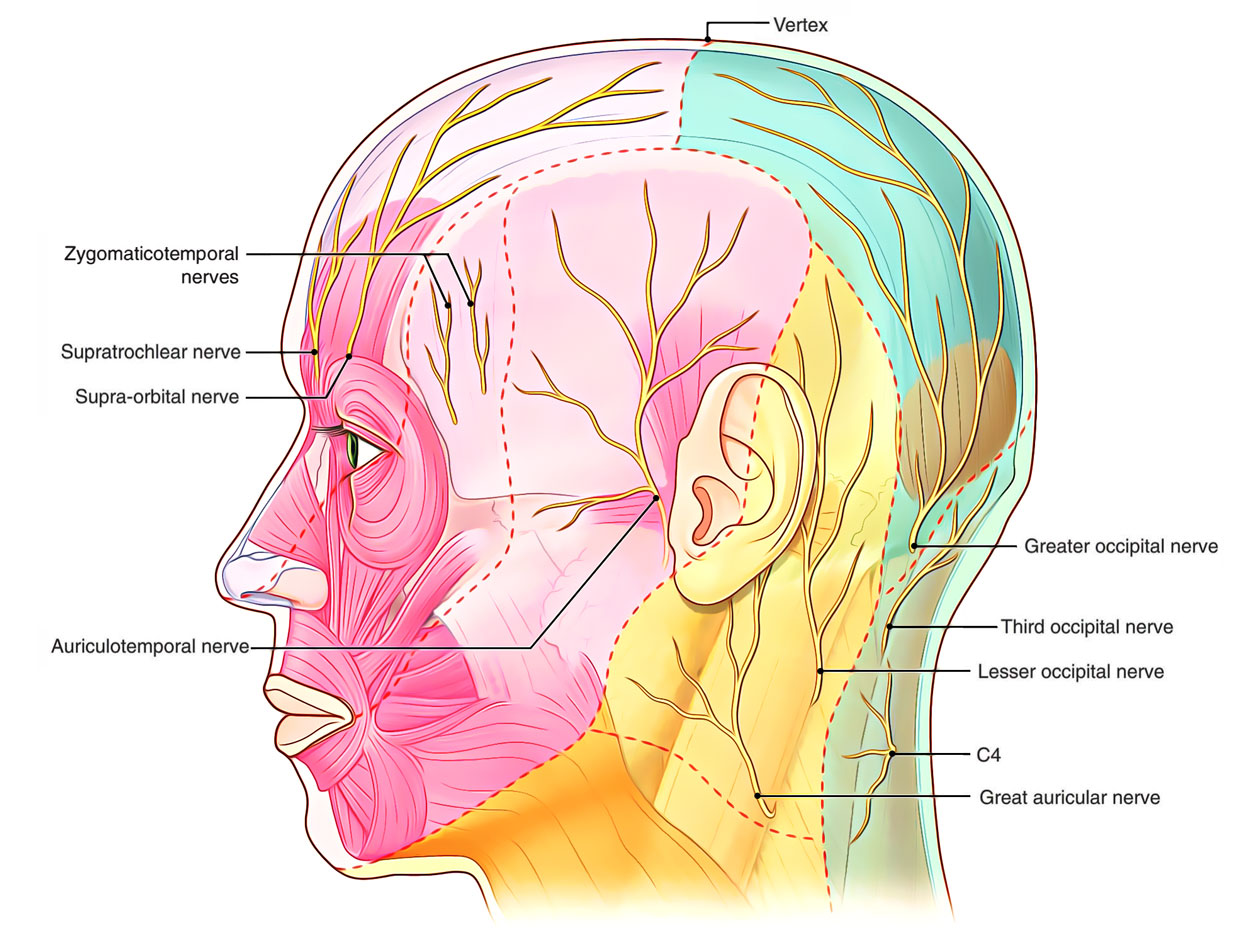
The scalp receives cutaneous innervation from branches of the trigeminal nerve (CN V) and cervical nerves (C2–C3).
- Trigeminal Nerve (CN V) Branches:
- Supratrochlear nerve – anteromedial forehead
- Supraorbital nerve – forehead to vertex
- Zygomaticotemporal nerve – temple
- Auriculotemporal nerve – area above the auricle
- Cervical Nerve Branches:
- Lesser occipital nerve (C2) – posterior to the ear
- Greater occipital nerve (C2) – occipital region
- Great auricular nerve (C2, C3) – skin posterior to ear and over mandibular angle
- Third occipital nerve (C3) – inferior occipital region
Clinical Notes & Key Points:
- Scalp Lacerations: Scalp wounds bleed profusely because tension by the occipitofrontalis muscle prevents vessel closure, dense connective tissue prevents vasoconstriction, and there is a rich anastomotic blood supply.
- Danger Area: The loose areolar layer is the danger area because infections can spread intracranially via valveless emissary veins.
- Layer Movement: The first three layers of the scalp move as a single unit.
- Innervation Split: Innervation comes from CN V (anterior scalp) and C2–C3 (posterior scalp).
You must be logged in to post a comment.