Learning Objectives
By the end of this session, the learner will be able to describe any articulation using a five-part systematic framework: Site, Type, Synovial Sub-classification, Actions, and Range of Motion (ROM). This structured approach is fundamental for diagnosing sports injuries, managing arthritis, and assessing physical rehabilitation.
The 5-Part Framework for Arthrology
To master the mechanics of the human frame, avoid memorizing joints as isolated facts. Instead, apply this rubric to understand the balance between stability and mobility.
1. Site (Anatomical Location)
Identifies the specific articulating bones and the body region. This provides the context for which muscle groups will cross the joint.
| Joint Name | Articulating Bones | Region |
|---|---|---|
| Elbow Joint | Humerus, Radius, and Ulna | Upper Limb (Arm/Forearm) |
| Hip Joint | Acetabulum (Pelvis) and Femur | Lower Limb (Trunk/Thigh) |
| Talocrural Joint | Tibia, Fibula, and Talus | Lower Limb (Leg/Foot) |
2. Type (Structural Classification)
Joints are classified by the material that binds the bones. This dictates the joint’s inherent stability and baseline mobility.
| Joint Type | Binding Material | Example |
|---|---|---|
| Fibrous | Dense connective tissue | Skull Sutures (Immobile) |
| Cartilaginous | Hyaline or Fibrocartilage | Intervertebral Discs |
| Synovial | Fluid-filled joint cavity | Knee / Shoulder (Highly mobile) |
Activity:
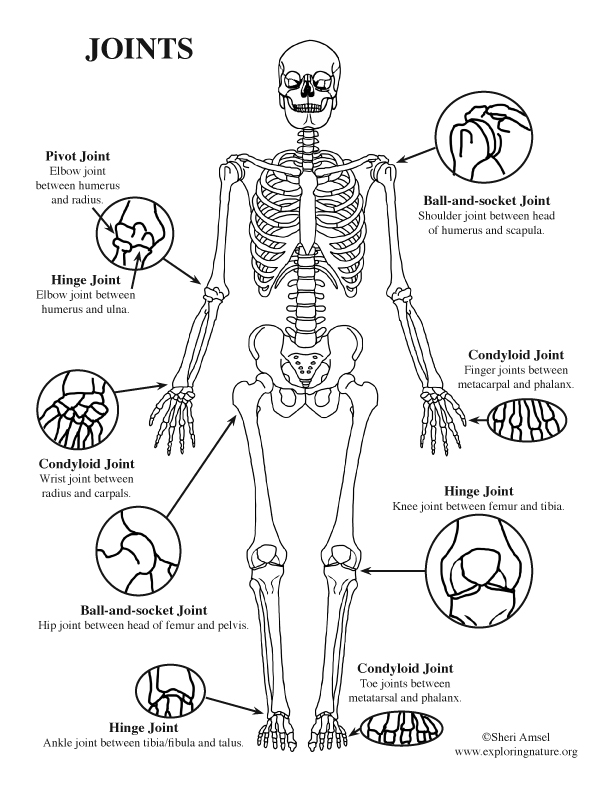
3. Types of Synovial Joints (Morphology)
Synovial joints are further sub-classified by the shape of the articulating surfaces, which defines the degrees of freedom.
| Shape | Permitted Movement | Example |
|---|---|---|
| Hinge | Uniaxial (Flexion/Extension) | Elbow |
| Ball and Socket | Multiaxial + Rotation | Hip / Shoulder |
| Saddle | Biaxial (Freedom in 2 planes) | Thumb CMC Joint |
| Pivot | Rotation only | Atlanto-axial joint |
4. Actions (Kinesiology)
The specific movements allowed. These are determined by the joint’s shape and the surrounding ligaments.
| Joint | Primary Actions |
|---|---|
| Hip | Flex/Ext, Abd/Add, Medial/Lateral Rotation. |
| Talocrural | Dorsiflexion, Plantarflexion. |
5. Range of Motion (ROM)
The measurable degree of movement. This is a critical metric for assessing recovery after injury or surgery.
| Joint Movement | Normal ROM (Approximate) | Clinical Tool |
|---|---|---|
| Hip Abduction | 45° | Goniometer |
| Elbow Flexion | 0° to 150° | Goniometer |
| Plantarflexion | 50° | Goniometer |
Activity:
Clinical Pearls for Medical Students:
- Stability vs. Mobility: The Shoulder is the most mobile joint, but also the most frequently dislocated. The Hip is structurally similar (Ball & Socket) but sacrifices mobility for the stability required for weight-bearing.
- Joint Capsules: In Synovial joints, the presence of a capsule means that infection (Septic Arthritis) or inflammation can cause significant pressure and pain within that closed space.
- Passive vs. Active ROM: If a patient has limited Active ROM but full Passive ROM, the problem is likely muscular or neurological. If both are limited, the pathology is likely within the joint itself.
You must be logged in to post a comment.