Learning Objectives
By the end of this session, the learner will be able to describe any visceral organ using a six-part systematic framework: Site, Shape, Size/Weight, Relations, Nerve Supply, and Blood Supply. This rubric is essential for physical examination, surgical approach, and interpreting diagnostic imaging.
The 6-Part Framework for Visceral Anatomy
To master the complex internal environment of the body, use this consistent rubric. It helps you organize clinical findings and understand how pathology in one organ can affect its neighbors.
1. Site (Topographical Region)
Identifies the anatomical cavity or quadrant where the organ resides. This is the first step in localizing referred pain.
| Organ | Primary Site | Clinical Localization |
|---|---|---|
| Liver | Abdomen | Right Upper Quadrant (RUQ). |
| Bladder | Pelvis | Retropubic space (when empty). |
| Eye | Head | Located within the bony orbit. |
2. Shape (Morphology)
The form of an organ often dictates its internal capacity and how it fits within a confined space.
| Shape Description | Organ Example | Functional Logic |
|---|---|---|
| Spherical | Eye | Optimized for rotation in multiple axes. |
| Bean-shaped | Kidney | Concave hilum for entry/exit of vessels. |
| Flat/Pear-shaped | Pancreas | Tucked behind the stomach for protection. |
3. Size / Weight (Physical Dimensions)
Physical parameters are vital for detecting organomegaly (enlargement) during percussion or palpation.
| Organ | Relative Scale | Clinical Importance |
|---|---|---|
| Liver | Large (~1.5 kg) | Weight varies with congestion (e.g., Heart Failure). |
| Thyroid | Small/Butterfly | Enlarges (Goiter) in iodine deficiency or autoimmunity. |
4. Relations (Spatial Anatomy)
Describes the organ’s neighbors and its relationship to the peritoneum. This is arguably the most important section for surgeons.
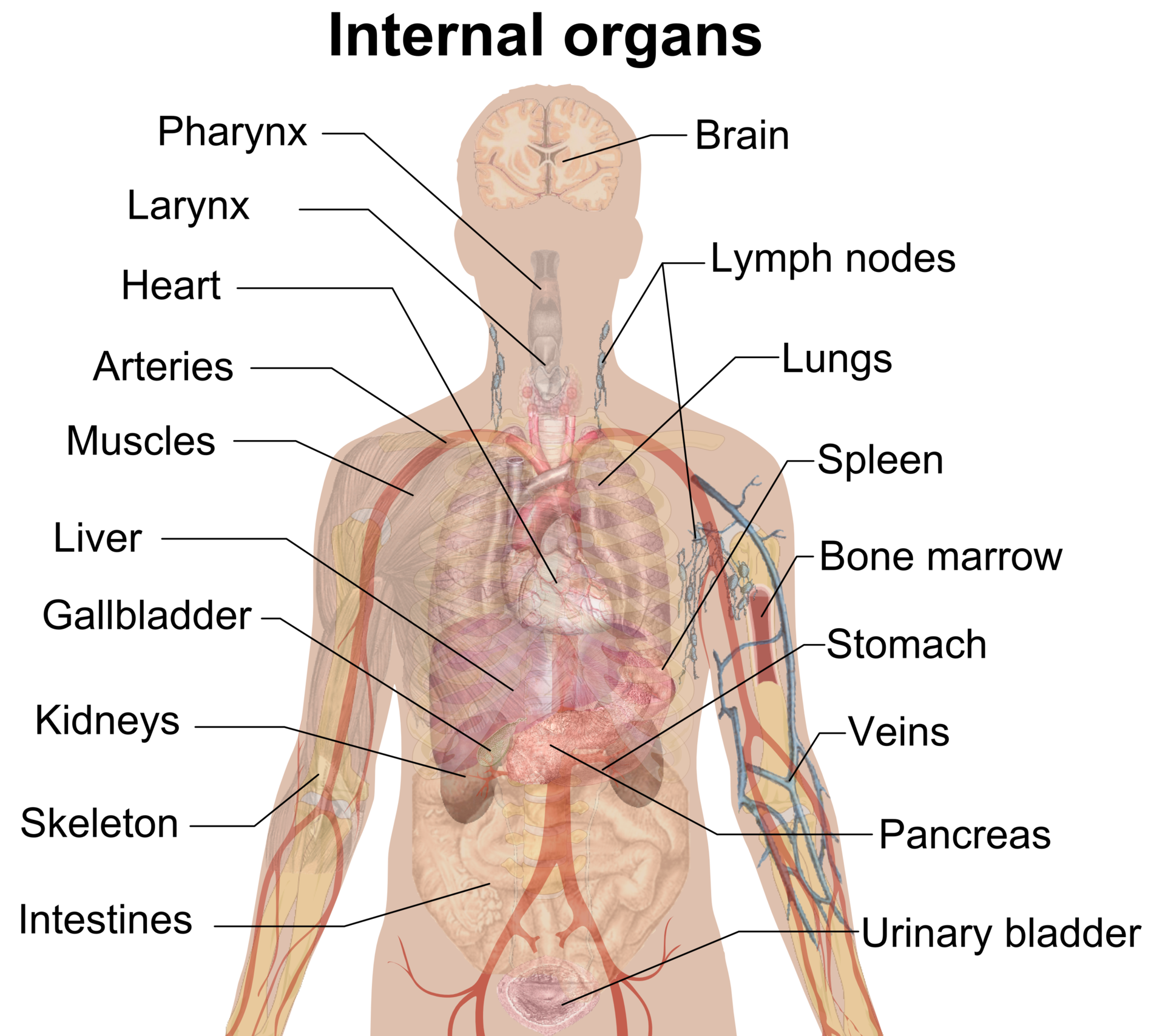
[Image showing the stomach and its relations to the pancreas and spleen]
| Type of Relation | Description | Example |
|---|---|---|
| Anterior/Posterior | What lies in front or behind? | The stomach is anterior to the pancreas. |
| Retroperitoneal | Behind the peritoneal sac. | Kidneys and Pancreas. |
| Intraperitoneal | Wrapped in peritoneum. | Spleen and Stomach. |
5. Nerve Supply (Innervation)
Most visceral organs have autonomic innervation (Sympathetic/Parasympathetic) to regulate involuntary functions.
| Nerve/Plexus | Target Organ | Function |
|---|---|---|
| Cardiac Plexus | Heart | Regulates heart rate and contractility. |
| Pelvic Splanchnic | Bladder | Parasympathetic: Contraction for voiding. |
| Hepatic Plexus | Liver | Regulates metabolic activity and bile flow. |
6. Blood Supply (Vasculature)
Describes the “life-line” of the organ. Understanding this is critical for diagnosing ischemic conditions (like a renal infarct).
| Arterial Supply | Venous Drainage | Target Organ |
|---|---|---|
| Renal Artery | Renal Vein | Kidney |
| Hepatic Artery / Portal Vein | Hepatic Veins | Liver |
| Superior Vesical a. | Vesical venous plexus | Bladder |
Activity:
Clinical Pearls for Medical Students:
- Referred Pain: Because of the Nerve Supply, visceral pain is often felt in a different location (e.g., Gallbladder pain felt in the right shoulder due to phrenic nerve irritation).
- Surgical “Windows”: The Relations section tells you which organs you have to move to get to another. To reach the pancreas, a surgeon often has to go through the “lesser sac” behind the stomach.
- Portal System: The liver is unique in its Blood Supply because it receives nutrient-rich blood from the Portal Vein and oxygen-rich blood from the Hepatic Artery.
You must be logged in to post a comment.