
Granulomatous Inflammation: High-Yield Pathology
Granulomatous inflammation is a distinctive, specialized pattern of chronic inflammation dominated by collections of activated macrophages that develop an elongated, epithelial-like appearance (epithelioid histiocytes). It represents a Type IV (delayed-type) hypersensitivity response mounted to wall off a persistent, poorly degradable offending agent.
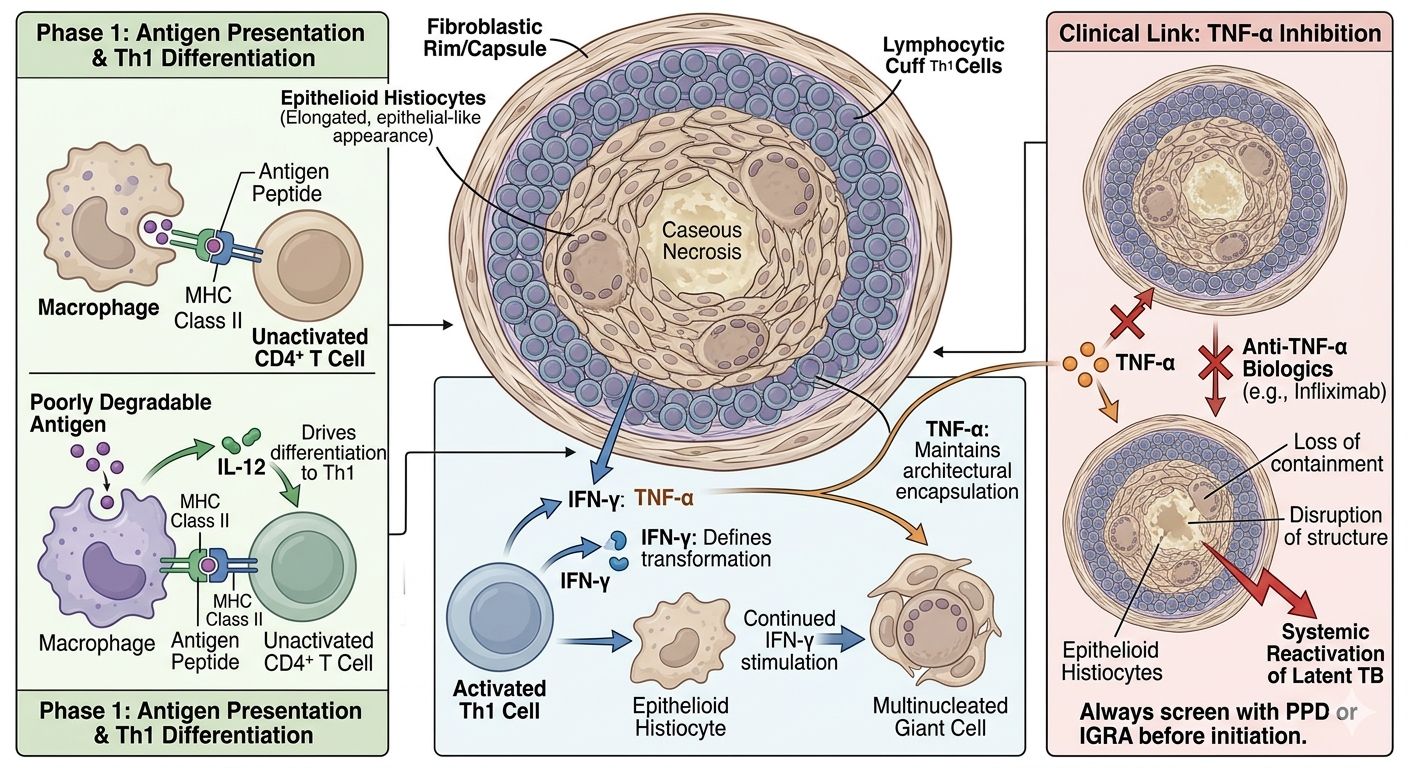
1. Cellular Architecture & Signaling Pathway
The coordinated orchestration of a granuloma depends entirely on a tightly regulated cytokine dialogue between macrophages and CD4+ T helper 1 (Th1) cells:
- Antigen Presentation: Macrophages or dendritic cells engulf the foreign material, processing and presenting it via MHC class II molecules to naive CD4+ T helper cells.
- Th1 Differentiation: Macrophages secrete IL-12, which drives the differentiation of naive T cells into antigen-specific Th1 cells.
- Macrophage Activation: Formed Th1 cells secrete Interferon-gamma (IFN-γ). This cytokine is the definitive molecular switch that transforms standard macrophages into highly microbicidal epithelioid histiocytes.
- Fusion & Containment: Under continued IFN-γ stimulation, epithelioid histiocytes fuse to form multinucleated giant cells. Macrophages also produce TNF-α, which maintains the structural integrity and architectural encapsulation of the granuloma.
2. Histological Components
Under light microscopy, a fully formed classic granuloma exhibits a highly organized, concentric layered layout from the center outwards:
- Central Core: Can be acellular, amorphous, and necrotic (caseating) or remain densely packed with intact cells (non-caseating).
- Epithelioid Histiocytes: Modified macrophages featuring abundant pink (eosinophilic) cytoplasm and elongated, pale, slipper-shaped or “shoe-sole” nuclei.
- Multinucleated Giant Cells:
- Langhans Giant Cells: Nuclei are systematically arranged along the periphery of the cell in a horseshoe-shaped or ring configuration. Highly characteristic of Mycobacterial infections.
- Foreign Body Giant Cells: Nuclei are disorganized and scattered randomly throughout the cytoplasm. Seen in response to non-immunogenic material (e.g., talc, sutures).
- Lymphocytic Rim: A peripheral defensive wall consisting of dark-staining Th1 lymphocytes, plasma cells, and an outermost zone of fibroblasts laying down protective collagen fibers.
3. Differential Diagnosis: Caseating vs. Non-Caseating
The classification of central necrosis serves as the primary diagnostic branch point utilized on pathology board examinations to categorize the underlying pathology:
| Type | Pathology & Mechanisms | Classic Clinical Examples |
|---|---|---|
| Caseating (Necrotizing) |
• Characterized by a central zone of cheesy, amorphous, acellular debris. • Driven by delayed-type hypersensitivity, causing severe macrophage-mediated free radical and hypoxic destruction of central cells. |
|
| Non-Caseating (Non-Necrotizing) |
• Lacks a central zone of acellular necrotic breakdown. • The internal core remains entirely populated by live, viable epithelioid histiocytes and multinucleated giant cells. |
|
4. Specific High-Yield Exam Variants
- Cat-Scratch Disease (Bartonella henselae): Characterized by highly unique stellate (star-shaped) necrotizing granulomas, typically localized within the regional draining lymph nodes (axillary, epitrochlear, or cervical).
- Leprosy (Mycobacterium leprae):
- Tuberculoid Leprosy: Strong host Th1 cell-mediated response yields well-formed, intact non-caseating granulomas that successfully restrict the bacilli, leading to localized, hypopigmented skin patches with asymmetric nerve damage.
- Lepromatous Leprosy: Weak host Th1 response (Th2 skewed) leads to a failure to form protective granulomas. Foamy macrophages become packed with massive quantities of acid-fast bacilli, leading to diffuse skin nodules and classic “leonine facies.”
You must be logged in to post a comment.