
Cellular Adaptations: High-Yield Pathology
Cellular adaptations are reversible structural or functional changes made by cells in response to physiological or pathological stress. When the limits of these adaptive responses are exceeded, or if the stress is inherently injurious, cell injury ensues.
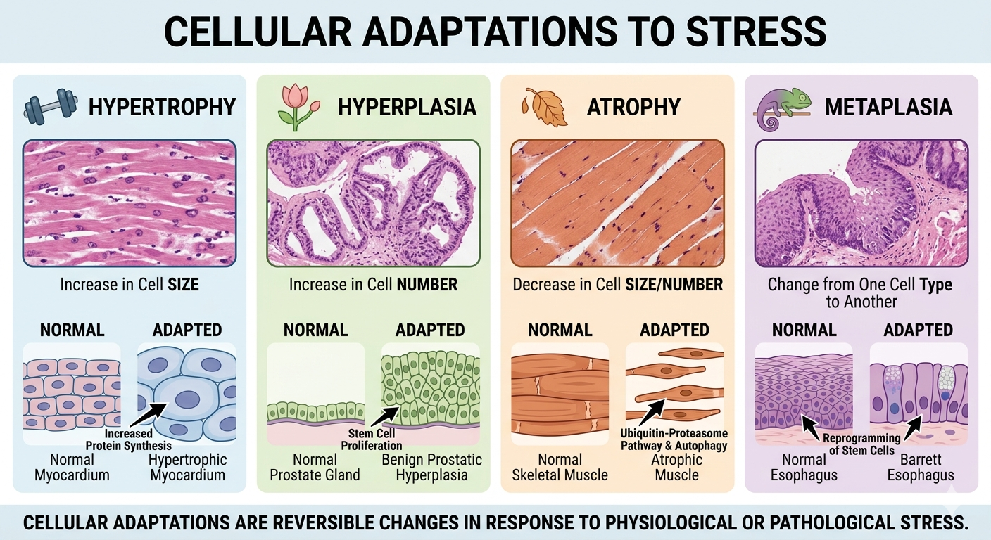
1. Core Mechanisms & Definitions
| Adaptation | Definition | Primary Mechanisms | Classic Board Examples |
|---|---|---|---|
| Hypertrophy | Increase in cell size, resulting in increased organ size. | Increased protein synthesis, gene activation, and production of structural filaments/organelles. Driven by mechanical stretch or growth factors. | • Left ventricular hypertrophy (LVH) due to systemic hypertension. • Uterine smooth muscle expansion during pregnancy. |
| Hyperplasia | Increase in cell number from stem cell proliferation. | Hormone- or growth factor-driven proliferation of mature cells or increased output from tissue stem cells. | • Benign Prostatic Hyperplasia (BPH). • Endometrial hyperplasia. • Compensatory liver regeneration post-partial resection. |
| Atrophy | Decrease in cell size and/or number, reducing organ size. | • Decreased size via the Ubiquitin-proteasome pathway (degrades the cytoskeleton) and autophagy. • Decreased number via apoptosis. |
• Skeletal muscle disuse (cast). • Loss of endocrine stimulation (postmenopausal endometrium). • Brain atrophy in Alzheimer’s disease. |
| Metaplasia | Reversible change where one differentiated cell type is replaced by another. | Reprogramming of local tissue stem cells or undifferentiated mesenchymal cells via transcription factor alterations, not transdifferentiation of mature cells. | • Barrett Esophagus: Squamous to columnar epithelium. • Squamous Metaplasia: Respiratory columnar to squamous epithelium in chronic smokers. |
2. Metaplasia Pathological Transitions
While metaplasia is an adaptive, reversible safety mechanism to handle chronic stress, it comes with a physiological trade-off (e.g., loss of mucus secretion or ciliary clearance in the respiratory tract) and can progress to malignancy if the noxious stimulus persists:
- Barrett’s Esophagus: Driven by chronic gastroesophageal reflux disease (GERD). The stratified squamous epithelium of the lower esophagus transitions to non-ciliated columnar epithelium with goblet cells (intestinal metaplasia) to withstand acidic conditions. This carries an increased risk of progressing to **Esophageal Adenocarcinoma**.
- Squamous Metaplasia of the Respiratory Tract: Driven by chronic cigarette smoking. The normal pseudostratified ciliated columnar epithelium transitions to stratified squamous epithelium to tolerate irritation. This increases the risk for **Squamous Cell Carcinoma** of the lung.
- Vitamin A (Retinol) Deficiency: Vitamin A is necessary for the proper differentiation of specialized epithelial surfaces (e.g., conjunctiva). Deficiency causes keratomalacia, where the normal thin, moist squamous lining of the eye undergoes metaplasia into a thickened stratum corneum, potentially causing blindness.
- Myositis Ossificans: A form of connective tissue metaplasia where intramuscular hematoma components differentiate into bone/cartilage following deep muscle trauma.
3. Hyperplasia vs. Neoplasia
Pathological hyperplasia (e.g., endometrial hyperplasia driven by unopposed estrogen exposure) creates a highly fertile soil for the accumulation of genetic mutations, increasing the risk of malignant conversion into neoplasia.
4. High-Yield Exam Pearls
- Benign Prostatic Hyperplasia (BPH): Exception to the rule. BPH is a pathological hyperplasia driven by dihydrotestosterone (DHT) binding to stromal and epithelial cells, yet it carries no increased risk for developing prostatic adenocarcinoma.
- Apoptosis vs. Atrophy: While tissue atrophy can involve apoptosis to reduce overall cell numbers, the characteristic visual shrinkage of individual cells during atrophy is mediated by the intracellular degradation of structural proteins via the ubiquitin-proteasome system.
You must be logged in to post a comment.