
Free Radical Injury & Oxidative Stress: High-Yield Pathology
Free radicals are highly reactive chemical species possessing a single unpaired electron in an outer orbital. Because of this unstable configuration, they aggressively initiate autocatalytic chain reactions, transferring their instability to adjacent cellular molecules and culminating in severe membrane, protein, and DNA damage.
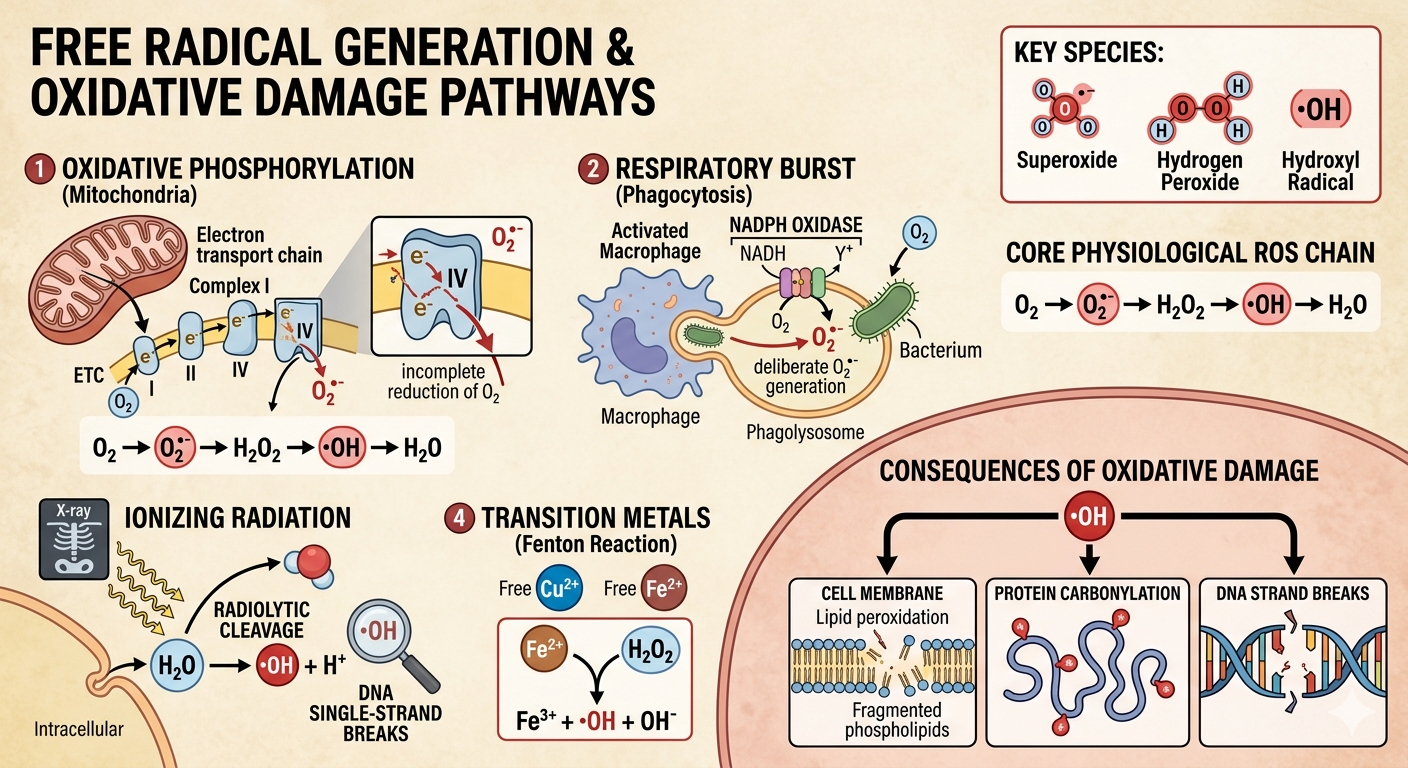
1. Generation of Reactive Oxygen Species (ROS)
Free radicals are produced physiologically during normal metabolic processes, but can spike dangerously under pathological states such as radiation, inflammation, or chemical toxicity:
- Oxidative Phosphorylation: Incomplete reduction of molecular oxygen (
) across the mitochondrial electron transport chain sequentially generates the core physiological ROS:
O₂ → O₂•⁻ (Superoxide) → H₂O₂ (Hydrogen Peroxide) → •OH (Hydroxyl Radical) → H₂O - Respiratory Burst (Phagocytosis): Activated neutrophils and macrophages deliberately synthesize ROS via NADPH Oxidase to destroy engulfed microbes, generating superoxide within the phagolysosome.
- Ionizing Radiation: Radiolytic cleavage of intracellular water (
) yields the highly toxic hydroxyl radical, which is the principal mediator of radiation-induced DNA strand breaks.
- Transition Metals: Free copper (
) or iron (
) participates directly in the **Fenton Reaction**, accelerating the conversion of hydrogen peroxide into the destructive hydroxyl radical:
Fe²⁺ + H₂O₂ → Fe³⁺ + •OH + OH⁻
2. Mechanisms of Cell Injury
Free radicals cause cellular collapse primarily through three highly destructive pathways:
| Target | Pathological Mechanism & Consequence |
|---|---|
| Lipid Peroxidation of Membranes |
Hydroxyl radicals attack the double bonds of polyunsaturated fatty acids in organelles and plasma membranes. This self-propagating reaction yields peroxides, destroying membrane fluidity and compromising structural integrity (e.g., mitochondrial swelling, lysosomal enzyme leak). |
| Protein Modifications | Radicals cause cross-linking, structural folding alterations, and oxidation of amino acid side chains. This directly damages structural architecture and inactivates vital metabolic enzymes. |
| DNA Fragmentation | Free radicals react directly with nuclear and mitochondrial thymine residues, inducing single- and double-strand DNA breaks. This triggers the activation of the p53 tumor suppressor pathway, driving the cell into **Apoptosis**. |
3. Neutralization & Antioxidant Defense Systems
Cells eliminate harmful free radicals utilizing both non-enzymatic scavengers and highly specific enzymatic breakdown systems:
- Superoxide Dismutase (SOD): Catalyzes the conversion of highly toxic superoxide into less toxic hydrogen peroxide:
2O₂•⁻ + 2H⁺ → H₂O₂ + O₂ - Glutathione Peroxidase: Located primarily within mitochondria and cytoplasm. It converts hydrogen peroxide and hydroxyl radicals into water while simultaneously converting reduced glutathione (GSH) into oxidized glutathione (GSSG).
- Catalase: Located inside peroxisomes. It converts hydrogen peroxide into water and molecular oxygen:
2H₂O₂ → 2H₂O + O₂ - Endogenous Scavengers: Vitamins A, C, and E, alongside metal carrier proteins (such as transferrin, ferritin, and ceruloplasmin), function by binding free transition metals to block Fenton-type chemistry.
4. Classic Clinical Vignette Examples
- Ischemia-Reperfusion Injury: Following restoration of blood flow to ischemic tissue (e.g., post-MI coronary stenting), the sudden influx of oxygen reacts with damaged, uncoupled mitochondrial chains. This causes a massive, acute spike in free radical production, compounding myocardial necrosis.
- Carbon Tetrachloride (
) Toxicity: Historically encountered in the dry-cleaning industry. The hepatic cytochrome P450 system metabolizes
). This drives severe lipid peroxidation of the hepatocyte rough endoplasmic reticulum, causing ribosome detachment, a loss of apolipoprotein synthesis, and a classic resultant fatty liver (steatosis) with centrilobular necrosis.
- Acetaminophen Overdose: Excess acetaminophen depletes hepatic glutathione pools. Accumulation of the reactive metabolite **NAPQI** then binds covalently to cellular proteins, inducing direct oxidative damage and massive hepatic necrosis.
You must be logged in to post a comment.