
Tumor Classification: Benign vs. Malignant Neoplasms
The clinical and pathological differentiation of tumors into benign and malignant states depends heavily on distinct cellular morphology, growth kinetics, local structural boundaries, and the capacity for distant metastasis.
1. Primary Biological and Macro-Structural Distinctions
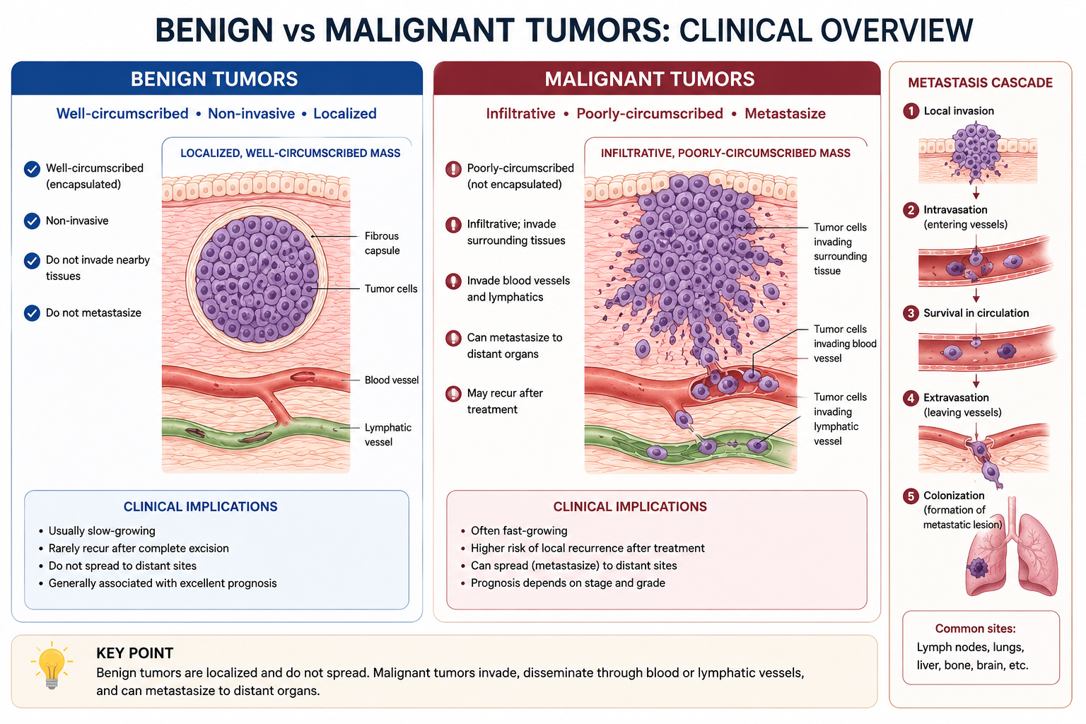
The definitive property that separates benign from malignant tumors is local tissue invasion and metastatic potential. While benign lesions grow as expansile, non-invasive masses, malignant lesions breach natural anatomical borders to invade local tissue and spread systemically.
2. Histopathological and Cytological Features
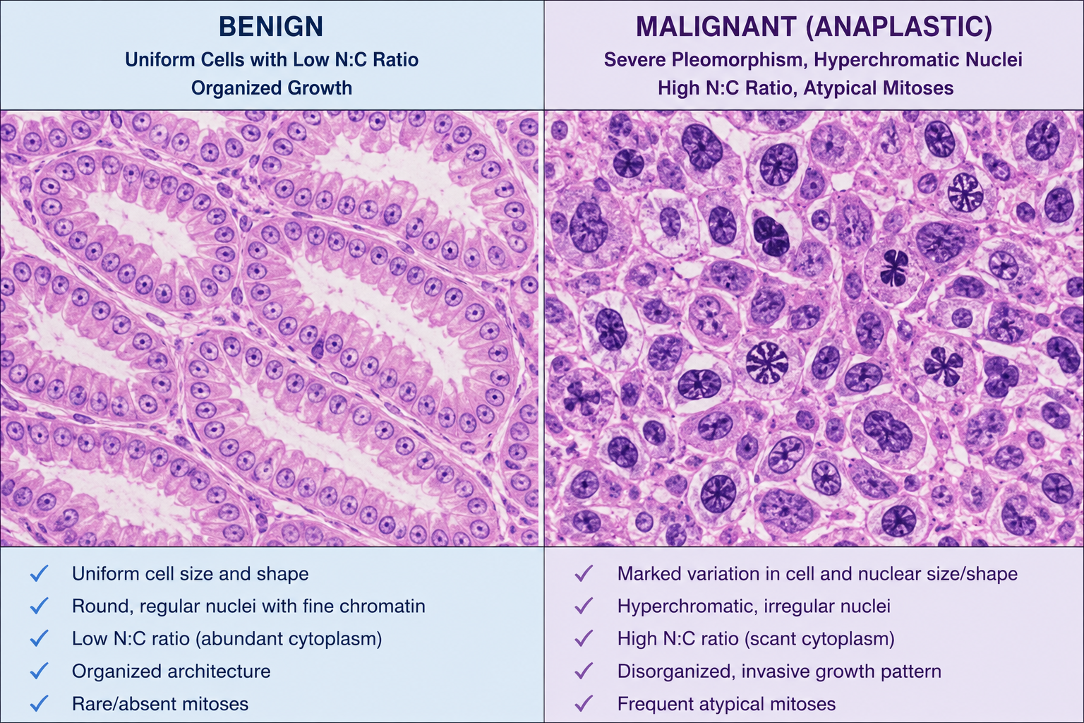
Evaluating standard hematoxylin and eosin (H&E) biopsy samples under high power reveals specific cytological details used to make an accurate diagnosis:
| Feature | Benign Neoplasms | Malignant Neoplasms |
|---|---|---|
| Differentiation | Well-differentiated: Cells closely resemble the normal architectural histology of the tissue of origin. | Poorly differentiated to anaplastic: Structural backward differentiation. Complete loss of structural orientation. |
| Growth Rate | Usually progressive and slow. Normal mitotic figures are rare. | Erratic and potentially rapid. Mitotic figures are numerous, prominent, and frequently atypical (e.g., tripolar spindles). |
| Local Invasion | Non-invasive: Cohesive, expansile growth. Often demarcated by a protective rim of fibrous tissue or a true capsule. | Invasive: Infiltrate, destroy, and systematically erode surrounding normal stromal borders. Lack a true capsule. |
| Metastasis | Absent: Never metastasize to regional or distant secondary organ sites. | Present: Frequently form secondary tumor colonies across distant organ systems via lymphatic, hematogenous, or serosal seeding. |
| Nuclear Morphology | Normal nuclear-to-cytoplasmic (N:C) ratio (approx. 1:4 or 1:6); uniform nuclear membrane. | High N:C ratio (up to 1:1): Large, irregular, hyperchromatic (darkly staining) nuclei with heavily clumped chromatin. |
| Cellular Pleomorphism | Minimal to absent variations in overall cellular size and shape. | Marked pleomorphism: Striking variation in individual cell sizes, custom shapes, and polarization. Giant cells are common. |
3. High-Yield Nomenclature Rules and Exceptions
While the suffix -oma paired with a tissue prefix usually points toward a benign growth configuration (e.g., fibroma, lipoma, chondroma, osteoma), there are multiple critical clinical exceptions to memorize for medical boards:
- Melanoma: Malignant tumor originating from cutaneous or mucosal melanocytes.
- Lymphoma: Solid malignant neoplasms derived from lymphoid lineage cells.
- Seminoma: Malignant germ cell tumor originating within the testicular germinal epithelium.
- Mesothelioma: Aggressive, asbestos-linked malignant neoplasm arising from mesothelial surface linings.
- Hepatoma: Historically used short-form name for Hepatocellular Carcinoma (fully malignant).
You must be logged in to post a comment.