Anatomy is the foundation of all medical knowledge. For NEET PG, it is not just about memorizing attachments or foramina; it is about understanding the structural basis of clinical practice. While the syllabus is vast, the exam pattern is predictable—if you know where to look.
To build a strong foundation, integrate free high-yield medical courses and practice questions from mymedschool.org into your daily routine. This platform is a fantastic resource for reinforcing core anatomical concepts through active recall.
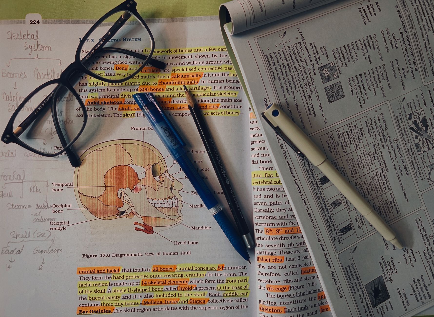
IN01 Anatomy 173
- Heart morphogenesis
- Heart embryology
- Fetal-postnatal derivatives
- Fetal circulation
- Heart anatomy
- Pericardium
- Coronary blood supply
- Thyroid development
- Pituitary gland
- Adrenal cortex and medulla
- Endocrine pancreas cell types
- Tongue development
- Normal gastrointestinal embryology
- Ventral wall defects
- Congenital umbilical hernia
- Tracheoesophageal anomalies
- Hypertrophic pyloric stenosis
- Intestinal atresia
- Pancreas and spleen embryology
- Retroperitoneal structures
- Important gastrointestinal ligaments
- Digestive tract anatomy
- Digestive tract histology
- Abdominal aorta and branches
- Nutcracker syndrome
- Superior mesenteric artery syndrome
- Gastrointestinal blood supply and innervation
- Celiac trunk
- Portosystemic anastomoses
- Pectinate line
- Liver tissue architecture
- Biliary structures
- Femoral region
- Inguinal canal
- Myopectineal orifice
- Hernias
- Fetal erythropoiesis
- Blood groups
- Hemolytic disease of the fetus and newborn
- Hematopoiesis
- Neutrophils
- Erythrocytes
- Thrombocytes (platelets)
- Monocytes
- Macrophages
- Dendritic cells
- Eosinophils
- Basophils
- Mast cells
- Lymphocytes
- Natural killer cells
- B cells
- T cells
- Plasma cells
- Immune system organs
- Lymph node
- Lymphatic drainage associations
- Spleen
- Thymus
- Upper extremity nerves
- Rotator cuff muscles
- Arm abduction
- Brachial plexus lesions
- Wrist region
- Hand muscles
- Distortions of the hand
- Knee exam
- Actions of hip muscles
- Lower extremity nerves
- Ankle sprains
- Signs of lumbosacral radiculopathy
- Neurovascular pairing
- Neural development
- Regionalization of the neural tube
- Central and peripheral nervous systems origins
- Neural tube defects
- Brain malformations
- Posterior fossa malformations
- Syringomyelia
- Kidney embryology
- Potter sequence
- Horseshoe kidney
- Congenital solitary functioning kidney
- Duplex collecting system
- Posterior urethral valves
- Vesicoureteral reflux
- Renal blood flow
- Glomerular anatomy
- Course of the ureters
- Early embryonic development
- Embryologic derivatives
- Teratogens
- Types of errors in morphogenesis
- Fetal alcohol syndrome
- Neonatal abstinence syndrome
- Placenta
- Amniotic fluid
- Twinning
- Twin-twin transfusion syndrome
- Umbilical cord
- Urachus
- Vitelline duct
- Pharyngeal apparatus
- Pharyngeal cleft derivatives
- Pharyngeal pouch derivatives
- Pharyngeal arch derivatives
- First and second pharyngeal arch syndromes
- Orofacial clefts
- Genital embryology
- Sexual differentiation
- Uterine (Müllerian duct) anomalies
- Male/female genital homologs
- Congenital penile abnormalities
- Descent of testes and ovaries
- Drainage of reproductive organs
- Female reproductive anatomy
- Adnexal torsion
- Pelvic organ prolapse
- Female reproductive epithelial histology
- Male reproductive anatomy
- Genitourinary trauma
- Autonomic innervation of male sexual response
- Seminiferous tubules
- Lung development
- Choanal atresia
- Lung malformations
- Club cells
- Alveolar cell types
- Neonatal respiratory distress syndrome
- Respiratory tree
- Lung anatomy
- Diaphragm structures
- Cells of the nervous system
- Sensory receptors
- Peripheral nerve
- Neuronal response to axonal injury
- Neurotransmitter changes with disease
- Meninges
- Blood-brain barrier
- Vomiting center
- Hypothalamus
- Thalamus
- Limbic system
- Cerebellum
- Basal ganglia
- Cerebral cortex regions
- Homunculus
- Cerebral arteries—cortical distribution
- Circle of Willis
- Dural venous sinuses
- Ventricular system
- Brainstem—ventral view
- Brainstem—dorsal view (cerebellum removed)
- Cranial nerve nuclei
- Vagal nuclei
- Brainstem cross sections- Midbrain
- Brainstem cross sections- Pons
- Brainstem cross sections- Medulla
- Cranial nerves and vascular pathways
- Cranial nerves and arteries
- Cranial nerves
- Cranial nerve reflexes
- Mastication muscles
- Spinal nerves
- Spinal cord—lower extent
- Conus medullaris and cauda equina syndrome
- Spinal cord levels and associated tracts
- Spinal tract anatomy and functions
- Landmark dermatomes
- Auditory anatomy
- Normal eye anatomy
- Aqueous humor pathway
- Ocular motility
Why Focus on These Topics?
The NEET PG exam uses Anatomy as a bridge to other subjects. A question on the Brachial Plexus is essentially a question on Orthopedics; a question on Cranial Nerves is a question on Neurology. By mastering these high-yield topics, you are simultaneously preparing for your clinical subjects, making your study time significantly more efficient.


High-Yield Anatomy Topics & Question Strategy
You must prioritize topics that frequently appear as clinical or image-based questions. For every topic you cover, test yourself immediately with questions to ensure the information sticks. (Brief Sample). List out a detailed outline.
| Region | Must-Master Topics | Why These? |
| Upper Limb | Brachial Plexus, Nerve Injuries (Erb’s, Klumpke’s, Wrist Drop) | High frequency in clinical scenarios. |
| Lower Limb | Sciatic Nerve, Femoral Triangle, Gait Abnormalities | Critical for Orthopedic surgery correlations. |
| Thorax | Coronary Circulation, Bronchopulmonary Segments | Essential for Cardiology & Pulmonology. |
| Abdomen | Inguinal Canal, Portosystemic Anastomosis, Hernias | Frequent surgical integration. |
| Neuroanatomy | Cranial Nerve Nuclei, Brainstem Lesions, Basal Ganglia | Complex but foundational for Neurology. |
| Head & Neck | Cavernous Sinus, Parotid Gland, Thyroid Arteries | Frequently tested in ENT and General Surgery. |
| Embryology | Pharyngeal Arches, Cardiac Development, GI Rotation | High “rattle-brain” potential; use diagrams! |
| Histology | Epithelium types, Lymph Node/Spleen structure | Common for image-based questions. |
Pro-Tips for Anatomy Mastery
1. Shift from Rote to Clinical
Stop trying to memorize every origin and insertion. Instead, ask yourself: “If this structure is injured, what will the patient look like?” If you understand that the radial nerve runs in the spiral groove, you automatically understand why a humerus fracture causes a wrist drop.
2. Visualize with Radiology
NEET PG is heavily reliant on image-based questions. Whenever you study a structure (like the thorax or abdomen), pull up a CT or MRI scan alongside your textbook. Identifying structures on an X-ray is a skill that must be practiced alongside your theory.
3. The Power of “Active Recall.”
Reading a textbook once is passive and often leads to quick forgetting. Use mymedschool.org to solve practice questions on these specific topics immediately after reading them. If you get a question wrong, revisit that specific paragraph. This is the fastest way to build long-term retention.
4. Create “High-Yield” Mnemonics
Anatomy is full of lists (contents of the cavernous sinus, branches of the carotid artery). Use mnemonics for these lists and keep them on sticky notes in your study area. Review these lists for 5 minutes every single morning.
5. Prioritize Previous Year Questions (PYQs)
The NBE loves recurring themes. Solve the last 5 years of PYQs to identify the “favorite” topics. If a specific nerve or artery has been asked about four times in the last decade, it is guaranteed to be a core focus for 2026.
Final Note: Anatomy is about pattern recognition. Don’t let the volume overwhelm you—break it down into these high-yield clusters, test yourself consistently, and link every structure to its clinical function.