Learning Objectives
Master the principles of Ventilation/Perfusion (
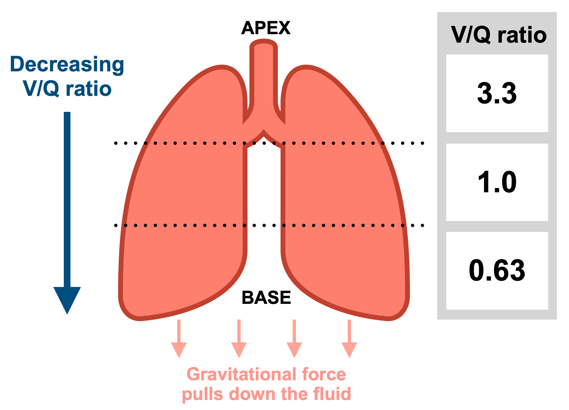
1. Regional Differences
Both ventilation (

| Region | Ratio |
Physiological State | Clinical Note |
|---|---|---|---|
| Apex (Zone 1) | ~3.0 (High) | Ventilation exceeds perfusion (“Wasted ventilation”). | High  favors Mycobacterium tuberculosis. favors Mycobacterium tuberculosis. |
| Middle (Zone 2) | ~0.8 – 1.0 | Ventilation and perfusion are best matched. | Ideal for gas exchange. |
| Base (Zone 3) | ~0.6 (Low) | Perfusion exceeds ventilation (“Wasted perfusion”). | Both and are at their absolute highest here. |
2. The Extremes: Shunt vs. Dead Space
When
| Scenario | Value |
Underlying Cause | 100% Response |
|---|---|---|---|
| Shunt | 0 | Airway obstruction (e.g., foreign body, pneumonia, pulmonary edema). | No improvement in  . . |
| Dead Space |  |
Blood flow obstruction (e.g., Pulmonary Embolism). | Improves (O2 reaches healthy alveoli). |
Activity:
3. Pressure Relationships (West Zones)
The relationship between Alveolar (


| Zone | Pressure Hierarchy | Mechanism |
|---|---|---|
| Zone 1 |  |
Alveolar pressure can collapse capillaries; minimal flow. |
| Zone 2 |  |
Flow is determined by the arterial-alveolar gradient. |
| Zone 3 |  |
Continuous flow; vessel pressures exceed alveolar pressure. |
Activity:
High-Yield Clinical Pearls:
- Exercise Recruitment: During exercise, increased cardiac output raises pulmonary artery pressure, opening apical capillaries. This makes the global
- The TB Connection: M. tuberculosis flourishes in the apex because that is where
is highest, providing a rich aerobic environment.
- The 100%
).
You must be logged in to post a comment.