
Learning Objectives
- Identify and describe the five secondary brain vesicles and their adult derivatives.
- Map the ventricular derivatives associated with each brain region.
- Apply developmental knowledge to understand congenital anomalies such as holoprosencephaly and anencephaly.
1. Primary Brain Vesicles
The embryonic neural tube forms the central nervous system (CNS) and undergoes regionalization to differentiate into three primary vesicles, which further divide into five secondary vesicles.
| Primary Vesicle | Region Derived | Future Structure / Secondary Vesicles |
|---|---|---|
| Prosencephalon (Forebrain) | Rostral end | Telencephalon and Diencephalon |
| Mesencephalon (Midbrain) | Middle portion | Mesencephalon (remains undivided) |
| Rhombencephalon (Hindbrain) | Caudal to midbrain | Metencephalon and Myelencephalon |
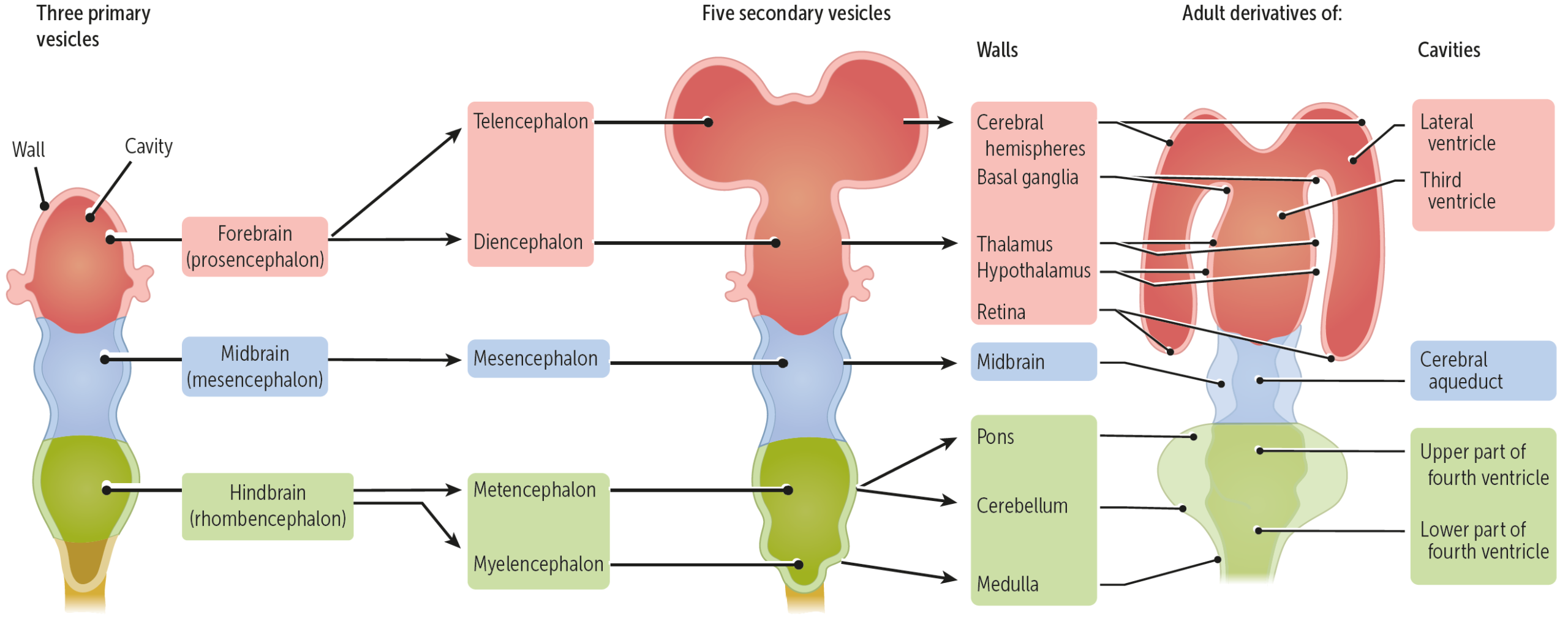
2. Secondary Brain Vesicles & Adult Derivatives
The cavities within each vesicle expand to form the ventricular system of the adult brain.
| Secondary Vesicle | Adult Brain Structure(s) | Ventricular Derivative |
|---|---|---|
| Telencephalon | Cerebral hemispheres | Lateral ventricles |
| Diencephalon | Thalamus, Hypothalamus | Third ventricle |
| Mesencephalon | Midbrain | Cerebral aqueduct |
| Metencephalon | Pons and Cerebellum | Upper part of the fourth ventricle |
| Myelencephalon | Medulla oblongata | Lower part of the fourth ventricle |
| Spinal Cord | Spinal cord proper | Central canal |
Clinical Notes & Correlations:
- Regionalization Defects: Failures or disruptions during early neural tube division can lead to major congenital anomalies such as holoprosencephaly and anencephaly.
Key Points & Memory Hooks:
CNS vs. PNS: The neural tube forms the entire CNS; the neural crest forms the PNS.
Development Sequence: The telencephalon develops first, followed by the diencephalon. Subsequent vesicles follow in alphabetical order (Mesencephalon, Metencephalon, Myelencephalon).
Ventricles: The cavity spaces of each secondary vesicle transform into the brain’s ventricular system.
CNS vs. PNS: The neural tube forms the entire CNS; the neural crest forms the PNS.
Development Sequence: The telencephalon develops first, followed by the diencephalon. Subsequent vesicles follow in alphabetical order (Mesencephalon, Metencephalon, Myelencephalon).
Ventricles: The cavity spaces of each secondary vesicle transform into the brain’s ventricular system.
You must be logged in to post a comment.