Learning Objectives
Differentiate between Pemphigus Vulgaris (PV) and Bullous Pemphigoid (BP) based on the molecular target (desmosomes vs. hemidesmosomes). Master the histological patterns (reticular vs. linear), clinical signs (Nikolsky sign), and the presence or absence of mucosal involvement.
1. Pathophysiology & Molecular Targets
Both conditions are Type II Hypersensitivity reactions involving IgG antibodies, but they attack different cellular “glues.”
| Condition | Antibody Target | Anatomical Location |
|---|---|---|
| Pemphigus Vulgaris | IgG vs. Desmoglein 1/3 (Desmosomes). | Intraepidermal (within the stratum spinosum). |
| Bullous Pemphigoid | IgG vs. BP180/BP230 (Hemidesmosomes). | Subepidermal (at the epidermal-dermal junction). |
2. Gross Morphology & Clinical Signs
The depth of the blister determines its stability and whether it affects the delicate mucosal membranes.
| Feature | Pemphigus Vulgaris (PV) | Bullous Pemphigoid (BP) |
|---|---|---|
| Blister Type | Flaccid (easy to rupture); intraepidermal. | Tense (firm); subepidermal. |
| Nikolsky Sign | Positive (skin sloughs with pressure). | Negative. |
| Mucosa | Commonly involved (oral ulcers). | Usually spared. |
| Histology | Acantholysis; “Row of tombstones”. | Subepidermal blister with Eosinophils. |
3. Immunofluorescence (IF) Patterns
Direct immunofluorescence reveals the specific “staining” pattern of the IgG antibodies on the biopsy sample.
| Condition | IF Pattern | Visual Description |
|---|---|---|
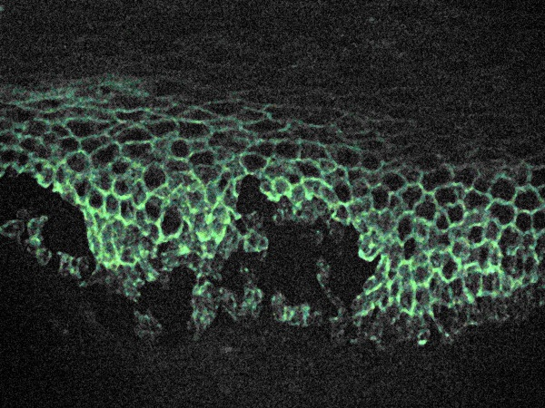
| Pemphigus Vulgaris | Reticular (Net-like) | Outlines individual keratinocytes like a fishnet.
|
| Bullous Pemphigoid | Linear | A smooth line at the epidermal-dermal junction.
|

Activity:
High-Yield Mnemonics:
- Bullous Pemphigoid: Antibodies are Bullow (Below) the epidermis at the Basement membrane.
- BP = Benign (less severe) + Brimming with Eosinophils.
- Pemphigus Vulgaris: Vulgaris is Very bad (potentially fatal) and involves the Vulva/Oral mucosa.
- Pemphigus = Positive Nikolsky.
You must be logged in to post a comment.