
Learning Objectives
- Identify the embryonic folding defects responsible for ventral wall defects (rostral, lateral, and caudal folds).
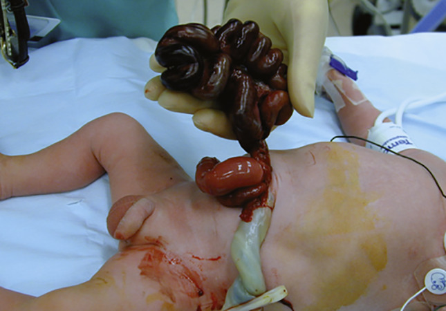
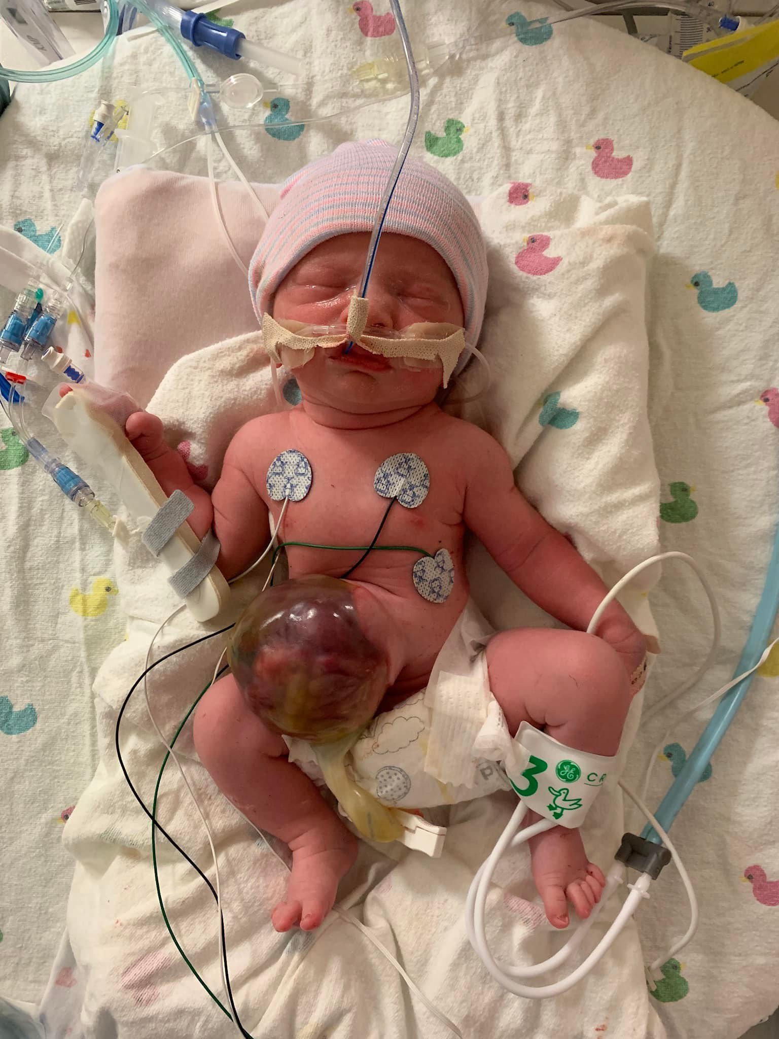
- Differentiate between gastroschisis and omphalocele based on presentation, peritoneal coverage, location, and associated anomalies.
- Recognize the clinical features and management of congenital umbilical hernias and their systemic associations.
1. Overview of Ventral Wall Defects
Ventral wall defects arise from the failure of embryonic folds to close properly during early development:
- Rostral Fold Closure Failure: Leads to sternal defects such as ectopia cordis.
- Lateral Fold Closure Failure: Leads to abdominal wall defects such as omphalocele and gastroschisis.
- Caudal Fold Closure Failure: Leads to anomalies such as bladder exstrophy.
2. Gastroschisis vs. Omphalocele
| Feature | Gastroschisis | Omphalocele |
|---|---|---|
| Presentation & Location | Paraumbilical herniation of abdominal contents (typically right-sided). | Herniation of abdominal contents through the umbilical ring (midline). |
| Sac Coverage | Not covered by peritoneum or amnion. | Covered by peritoneum and amnion (light gray, shiny sac). |
| Associations & Prognosis | Not commonly associated with chromosomal abnormalities; generally good prognosis.
|
Frequently associated with congenital anomalies (e.g., Trisomies 13 and 18, Beckwith-Wiedemann syndrome) and cardiac, GU, or neural tube defects.
|
3. Congenital Umbilical Hernia
- Definition: Caused by delayed closure of the umbilical ring following physiologic herniation of the midgut, leaving a patent umbilical orifice.
- Clinical Findings: Covered by skin; often reducible, but protrudes with increased intra-abdominal pressure (e.g., crying).
- Associations: May be linked to congenital disorders such as Down syndrome and congenital hypothyroidism.
- Outcome: Small defects usually close spontaneously.
Clinical Notes & Pearls:
- Sac Distinction: Remember that Gastroschisis has Gut exposed (No sac), whereas Omphalocele is enclosed in an Outer sac.
- Syndromic Check: An omphalocele requires a thorough workup for chromosomal abnormalities and cardiac anomalies, unlike isolated gastroschisis.
Key Points & Memory Hooks:
Fold Defects: Rostral = sternal/ectopia cordis; Lateral = body wall/omphalocele & gastroschisis; Caudal = bladder exstrophy.
Coverage: Omphalocele = Sac present (midline); Gastroschisis = No sac (right of umbilicus).
Umbilical Hernia: Skin-covered protrusion through the ring that typically resolves without intervention.
Fold Defects: Rostral = sternal/ectopia cordis; Lateral = body wall/omphalocele & gastroschisis; Caudal = bladder exstrophy.
Coverage: Omphalocele = Sac present (midline); Gastroschisis = No sac (right of umbilicus).
Umbilical Hernia: Skin-covered protrusion through the ring that typically resolves without intervention.
You must be logged in to post a comment.