
The Reverse Lordotic View
1. Clinical Definition
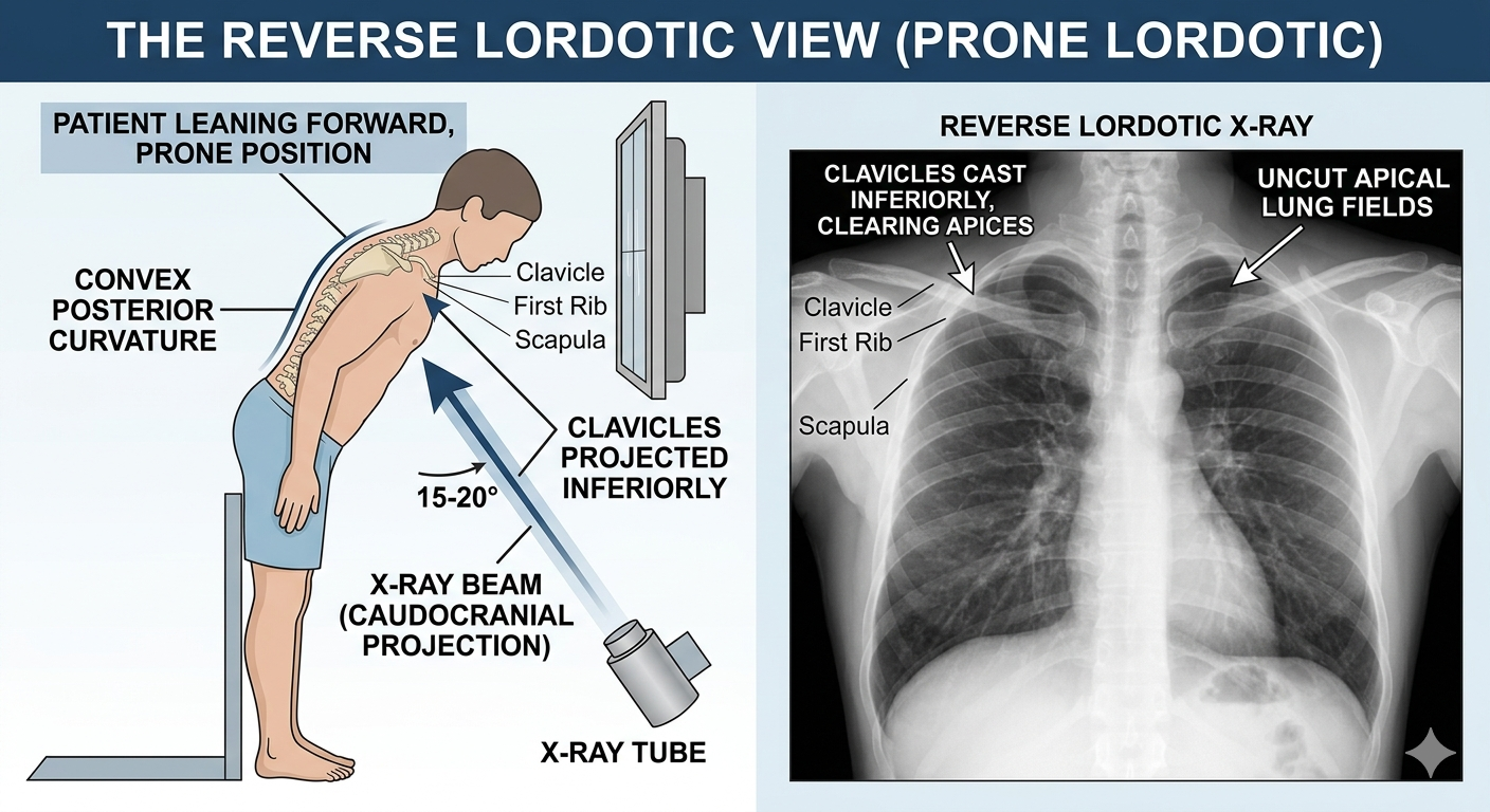
The Reverse Lordotic View (also known as the prone lordotic view or caudocranial projection) is a specialized chest X-ray view where the patient is positioned to lean forward with the X-ray beam angled cranially. This projects the clavicles inferiorly, away from the lung apices.
2. High-Yield Indications
- Obscured Apices: Used when a standard apical lordotic view is contraindicated, painful, or technically difficult for the patient.
- Clarification of Apical Lesions: Intended to clear the clavicles and first ribs from the lung apices to identify occult lesions, cavitary disease (e.g., TB), or apical scarring.
3. Key Differences
| Feature | Lordotic (Standard) | Reverse Lordotic |
|---|---|---|
| Patient Position | Leaning backward (supine). | Leaning forward (prone). |
| Primary Goal | Clear clavicles from apices. | Clear clavicles from apices. |
4. Exam Pearls
- Clinical Context: Like the classic lordotic view, this is primarily an historical/board-specific technique. It is physically demanding for patients and provides significantly less diagnostic information than a Chest CT.
- Board Strategy: If a question asks for a way to clear the clavicles from the lung apices and gives options, both lordotic and reverse lordotic views are functional equivalents for that specific diagnostic goal.
You must be logged in to post a comment.