
Necrosis vs. Apoptosis
Understanding the fundamental differences between these two modes of cell death is essential for pathology board examinations.
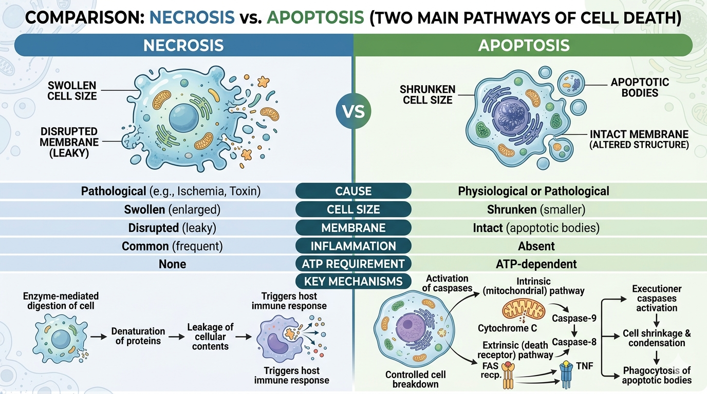
1. Comparative Table
| Feature | Necrosis | Apoptosis |
|---|---|---|
| Cause | Pathological (Ischemia, Toxin) | Physiological or Pathological |
| Cell Size | Swollen (enlarged) | Shrunken (smaller) |
| Membrane | Disrupted (leaky) | Intact (altered structure) |
| Inflammation | Common (frequent) | Absent |
| ATP Requirement | None | ATP-dependent |
2. Key Mechanisms
- Necrosis: Results from enzyme-mediated digestion of the cell, denaturation of proteins, and leakage of cellular contents, which triggers the host immune response.
- Apoptosis: A highly regulated “programmed” cell death. It involves the activation of caspases, leading to controlled cell suicide without causing damage to surrounding tissue. It can occur via the Intrinsic (mitochondrial) pathway (Cytochrome C release) or the Extrinsic (death receptor) pathway (FAS/TNF).
3. Exam Focus
- DNA Laddering: A classic feature of apoptosis where nucleases cleave DNA into fragments of 180–200 base pairs, visible as a ladder on gel electrophoresis.
- Necrosis Types: Always associate necrosis with tissue inflammation, whereas apoptosis is a “quiet” process often used for removing damaged or unwanted cells during development.
You must be logged in to post a comment.