
Learning Objectives
- Explain the hierarchical structural organization of DNA—from torsional supercoiling in circular genomes to eukaryotic nucleosomes, 30-nm chromatin fibers, looped domains, and metaphase chromosomes.
- Differentiate negatively supercoiled DNA (underwound, biologically active) from positively supercoiled DNA (overwound), and compare the enzymatic mechanisms and clinical pharmacology of Topoisomerase I and Topoisomerase II (DNA gyrase).
- Describe the protein composition of the nucleosome core octamer ($\text{2} \times [\text{H2A, H2B, H3, H4}]$) and the role of linker histone H1 in chromatin compaction.
- Contrast euchromatin (lightly packed, transcriptionally active) with heterochromatin (densely packed, transcriptionally silent) and explain the clinical utility of metaphase chromosomes in cytogenetic karyotyping.
To fit meters of linear genetic material inside microscopic cellular compartments without tangling—while still permitting rapid enzymatic access for replication, transcription, and repair—cells organize DNA into multi-tiered structural hierarchies ranging from topological supercoils to condensed mitotic chromosomes.
DNA Supercoiling & Topoisomerase Enzymes
Mitochondrial DNA and most prokaryotic chromosomes exist as closed circular DNA molecules whose topological state fluctuates between relaxed and supercoiled conformations depending on torsional strain.
| Topological State / Enzyme | Structural & Enzymatic Mechanism | Biological & Clinical Relevance |
|---|---|---|
| Negatively Supercoiled DNA | Forms when the DNA double helix is underwound relative to its relaxed B-DNA state. | Predominant, biologically active form; stores free energy that facilitates local strand separation required for replication and transcription. |
| Positively Supercoiled DNA | Forms when the DNA double helix is overwound (e.g., ahead of advancing replication forks). | Tightly wound and less favorable for biological reactions; must be relieved to prevent replication fork arrest. |
| Topoisomerase I | Creates transient single-stranded DNA breaks (nicks) to relieve torsional supercoils without requiring ATP. | Targeted by camptothecin antineoplastic analogs (irinotecan, topotecan) used in colorectal and ovarian cancers. |
| Topoisomerase II (e.g., Bacterial DNA Gyrase) |
Creates transient double-stranded DNA breaks in an ATP-dependent manner, introducing negative supercoils and untangling interlocked daughter circles. | • Prokaryotic DNA gyrase is inhibited by fluoroquinolones (ciprofloxacin, levofloxacin). • Eukaryotic Topoisomerase II is inhibited by etoposide, teniposide, and anthracyclines (doxorubicin). |
Activity
Nucleosomes, Chromatin & Higher-Order Packaging
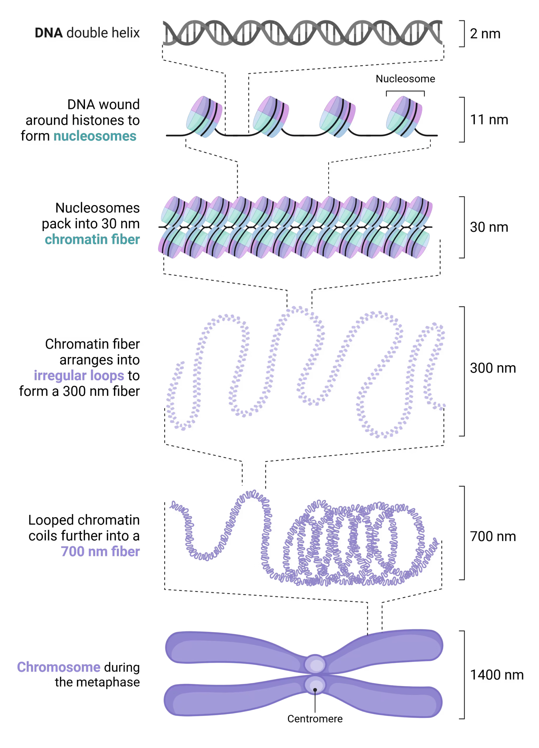
Hierarchical Levels of Eukaryotic DNA Compaction
- Histone Proteins: Small, evolutionarily conserved basic proteins enriched in positively charged amino acids—specifically lysine and arginine—that bind tightly via ionic interactions to the negatively charged sugar-phosphate backbone of DNA.
- Nucleosome Core Particle (10-nm Fiber): Approximately 147 base pairs (bp) of dsDNA wrap ~1.65 times around a core histone octamer composed of two copies each of four core histones: $2 \times (\text{H2A, H2B, H3, H4})$, forming the classic “beads on a string” 10-nm fiber.
- 30-nm Chromatin Fiber (Solenoid): Histone H1 binds to the linker DNA segments between adjacent nucleosomes, pulling nucleosomes together to compact the 10-nm fiber into a dense 30-nm solenoid fiber.
- Higher-Order Chromosome Architecture: The 30-nm fibers organize into radial looped domains anchored to non-histone nuclear scaffolding proteins, which further supercoil during prophase/metaphase into maximally condensed mitotic chromosome arms.
Euchromatin vs. Heterochromatin & Cell-Cycle Chromosomes
| Chromatin State | Structural Packaging & Staining | Transcriptional Activity & Genomic Locations |
|---|---|---|
|
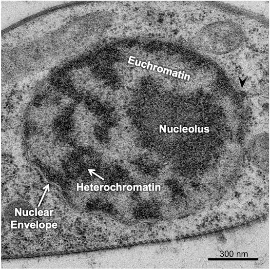
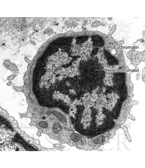
Euchromatin |
Lightly packed, decondensed, and extended chromatin fibers (appears pale/electron-lucent under microscopy). | Transcriptionally active; open steric configuration allows RNA polymerases and transcription factors free access to gene promoters. |
|
Heterochromatin
|
Densely packed, tightly coiled chromatin fibers (stains darkly/electron-dense; often associated with DNA methylation and histone deacetylation). | Transcriptionally silent; localized to centromeres, telomeres, and permanently inactivated regions such as the female inactivated X chromosome (Barr body). |
Activity
Chromosomes Across the Cell Cycle & Cytogenetic Karyotyping
- Interphase Nucleus
You must be logged in to post a comment.