
Learning Objectives
- Describe the external and internal anatomy of the midbrain (mesencephalon), including the tectum, cerebral peduncles, and internal nuclei.
- Differentiate between structural cross-sections at the level of the inferior colliculus versus the superior colliculus.
- Identify major cranial nerve emergences (CN III and CN IV) and fiber tracts traversing the midbrain.
- Examine the arterial blood supply originating primarily from branches of the basilar and posterior cerebral arteries.
1. External Anatomy & Landmarks
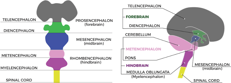
The midbrain is the most superior segment of the brainstem, measuring approximately 2 cm in length and linking the forebrain to the pons and cerebellum.
| Region | Key Structural Components | Functional Description |
|---|---|---|
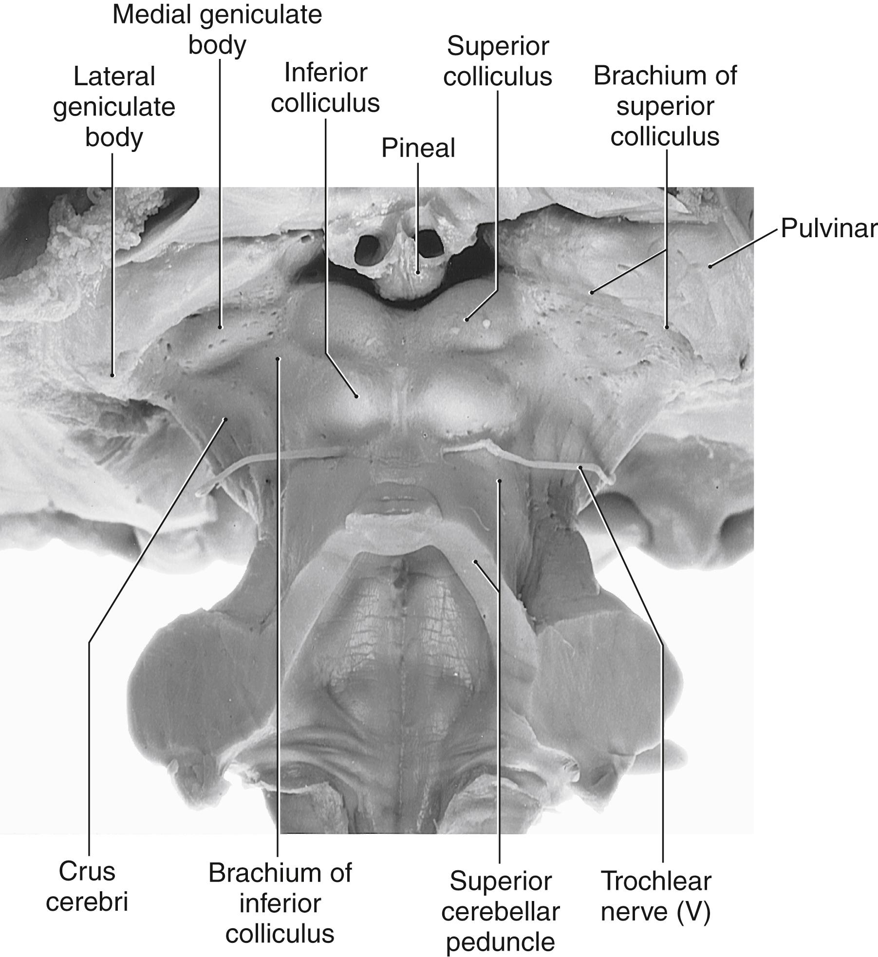
| Tectum (Posterior) | Contains the corpora quadrigemina (superior and inferior colliculi) and associated brachia. | Superior colliculi manage visual reflexes; inferior colliculi handle auditory relay. |
| Cerebral Peduncles (Anterior/Lateral) | Separated by the interpeduncular fossa containing the posterior perforated substance. | Anchor the cerebral hemispheres to the pons and transmit massive descending motor pathways. |
| Cranial Nerves | CN III (exits between peduncles) and CN IV (emerges posteriorly just below the inferior colliculi). | Provides crucial oculomotor (CN III) and trochlear (CN IV) innervation to extraocular muscles. |
2. Internal Anatomy & Transverse Levels
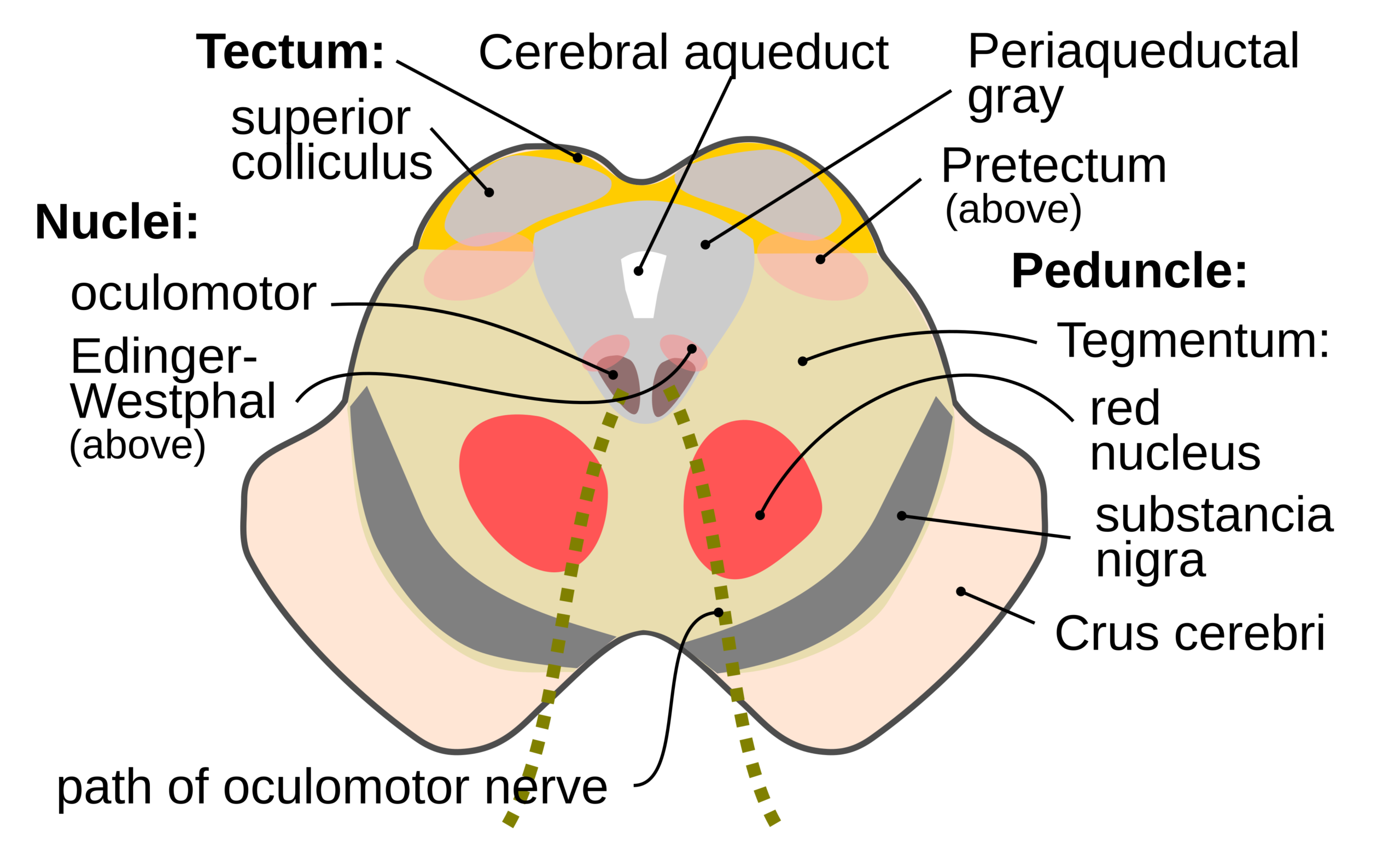
- Level of the Inferior Colliculus: Features the crus cerebri carrying descending tracts (corticospinal, corticobulbar, frontopontine, temporopontine), the substantia nigra (divided into pars compacta and pars reticulata), periaqueductal gray, trochlear nucleus (CN IV), and the decussation of the superior cerebellar peduncles.
- Level of the Superior Colliculus: Characterized by prominent paired red nuclei involved in motor coordination, the oculomotor nucleus (CN III replacing CN IV), and the rubrospinal tract decussation.
Vasculature & Clinical Notes:
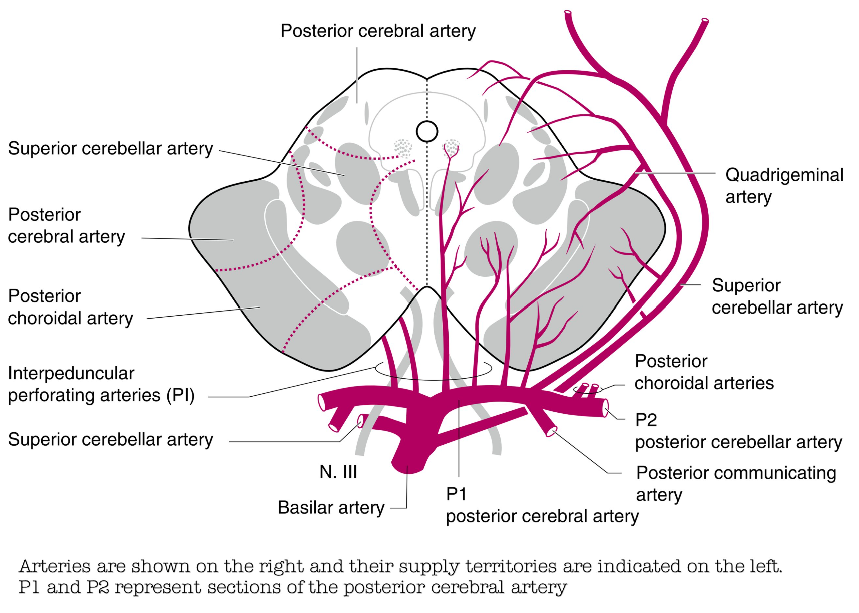
- Arterial Supply: Blood is supplied predominantly by branches of the basilar artery and posterior cerebral artery (PCA), including peduncular branches, superior cerebellar arteries, and interpeduncular perforating vessels.
- Midbrain Syndromes: Vascular occlusions or mass effects can damage local fascicles and nuclei, resulting in classic crossed brainstem paralyses (e.g., Weber’s syndrome involving CN III palsy alongside contralateral hemiplegia).
Key Points & Memory Hooks:
Colliculi Rule: Superior is for sight (you look *up* with superior eyes); Inferior is for sound.
Cranial Nerve Exit: CN III exits out the *front* between the peduncles; CN IV is unique as the only cranial nerve wrapping around to exit out the *back*.
Substantia Nigra Divider: The dark, dopaminergic substantia nigra acts as the dividing frontier separating the anterior crus cerebri from the posterior tegmentum.
Colliculi Rule: Superior is for sight (you look *up* with superior eyes); Inferior is for sound.
Cranial Nerve Exit: CN III exits out the *front* between the peduncles; CN IV is unique as the only cranial nerve wrapping around to exit out the *back*.
Substantia Nigra Divider: The dark, dopaminergic substantia nigra acts as the dividing frontier separating the anterior crus cerebri from the posterior tegmentum.
You must be logged in to post a comment.