
The Apical Lordotic View (Chest Radiography)
1. Clinical Definition
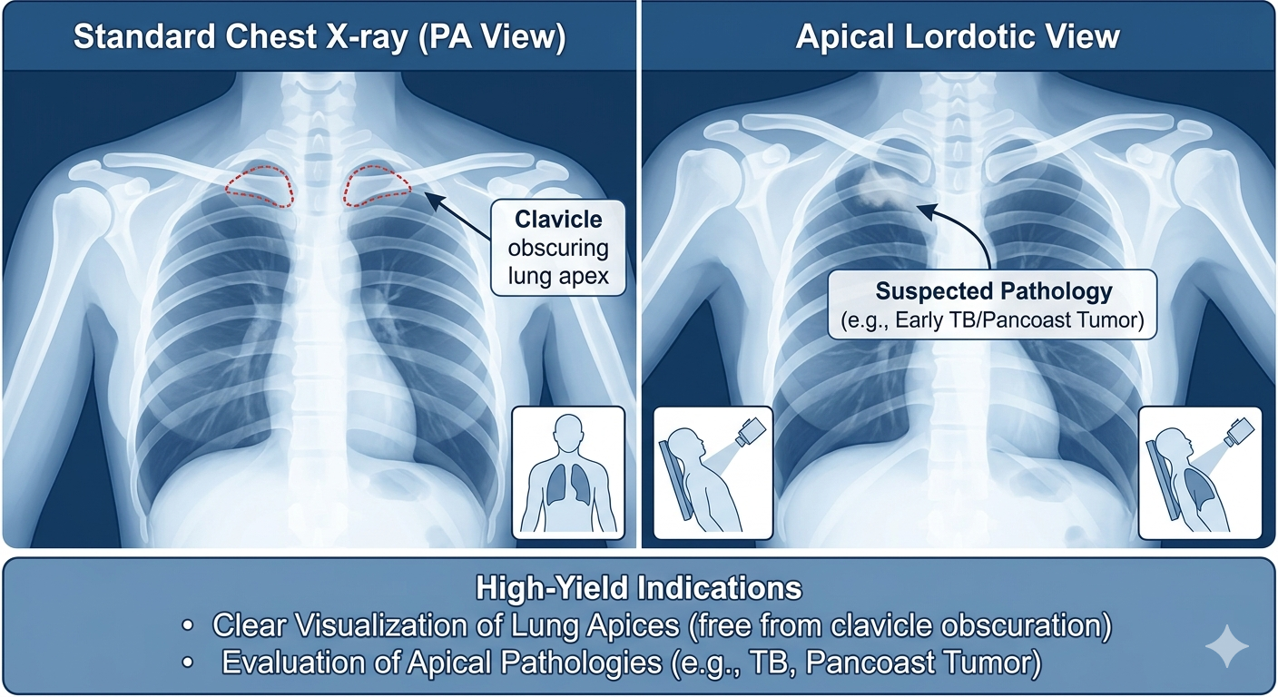
The Apical Lordotic View is a specialized chest X-ray projection where the patient is positioned to lean backward, projecting the clavicles superiorly and away from the lung apices. This maneuver “unfolds” the apex of the lungs.
2. High-Yield Indications
- Apical Pathologies: The primary goal is to visualize the lung apices clearly without obscuration by the clavicles and first/second ribs.
- Tuberculosis (TB): Historically, the most common indication, as pulmonary TB classically affects the apical and posterior segments of the upper lobes.
- Pancoast Tumor (Superior Sulcus Tumor): Useful for early evaluation of tumors at the apex of the lung.
3. Key Radiological Findings
| Pathology | Radiographic Goal |
|---|---|
| Apical TB | To see cavitary lesions or infiltrates hidden behind the clavicle. |
| Apical Pleural Cap | Evaluation of thickening or masses in the superior sulcus. |
4. Exam Pearls
- Exam Classic: If a board question presents a patient with chronic cough, night sweats, or hemoptysis and mentions an apical lesion partially obscured by the clavicles on a standard PA view, the next best imaging step to clarify the apex is the Apical Lordotic view.
- Limitations: Like other special views, this has been largely replaced by CT of the chest, which provides superior detail of the apices without the need for complex patient positioning.
You must be logged in to post a comment.