
Gell and Coombs Classification of Hypersensitivity
Hypersensitivity reactions are exaggerated or inappropriate immune responses to antigens, causing tissue damage rather than protection. These are categorized into four distinct types (Type I-IV) based on the immunological mechanism involved.
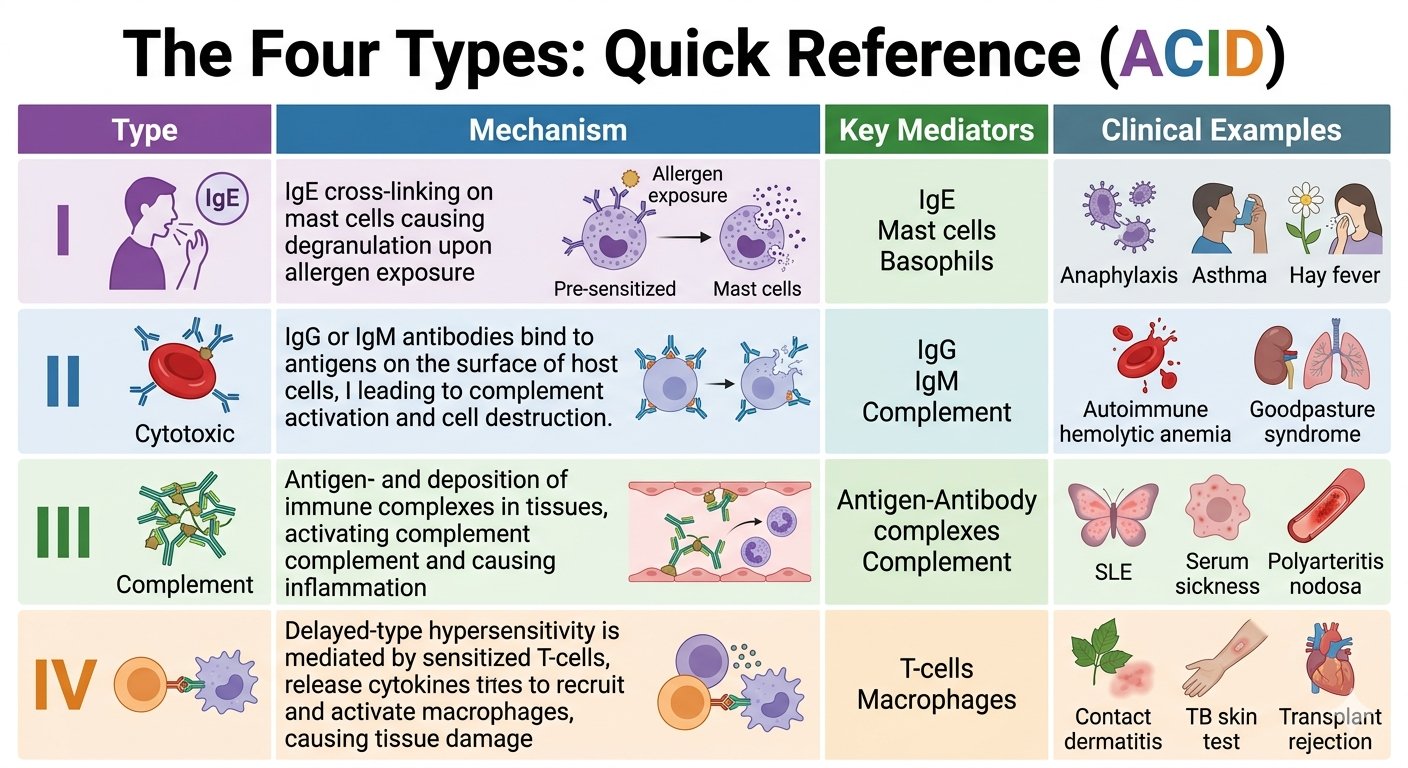
The Four Types: Quick Reference (ACID)
| Type | Mechanism | Key Mediators | Clinical Examples |
|---|---|---|---|
| I | Anaphylactic/Atopic | IgE, Mast cells, Basophils | Anaphylaxis, Asthma, Hay fever |
| II | Cytotoxic (Antibody-mediated) | IgG, IgM, Complement | Autoimmune hemolytic anemia, Goodpasture |
| III | Immune Complex | Ag-Ab complexes, Complement | SLE, Serum sickness, Polyarteritis nodosa |
| IV | Delayed-type | T-cells, Macrophages | Contact dermatitis, TB skin test, Transplant rejection |
Detailed Mechanisms
Type I: Immediate Hypersensitivity
Occurs within minutes of antigen exposure in a sensitized individual. Initial exposure induces class switching of B cells to IgE production. IgE binds to Fc receptors on mast cells and basophils. Subsequent exposure leads to cross-linking of IgE, causing degranulation and release of histamine, tryptase, and leukotrienes.
Type II: Antibody-Mediated Hypersensitivity
Antibodies (IgG or IgM) bind to antigens on cell surfaces or extracellular matrix. Damage occurs via three pathways:
- Opsonization/Phagocytosis: Antibody/complement-coated cells are cleared by macrophages.
- Complement-mediated lysis: Formation of the Membrane Attack Complex (MAC).
- Antibody-Dependent Cellular Cytotoxicity (ADCC): Natural Killer cells bind Fc receptors and induce apoptosis.
Type III: Immune Complex-Mediated Hypersensitivity
Antigen-antibody complexes are formed in circulation and subsequently deposited in tissues (vessels, joints, glomeruli). The deposition triggers complement activation, which attracts neutrophils. Neutrophils release lysosomal enzymes and reactive oxygen species, causing local tissue damage (vasculitis).
Type IV: Delayed-Type Hypersensitivity
The only type not mediated by antibodies. Driven by sensitized T cells (CD4+ or CD8+). Upon re-exposure, Th1 cells release cytokines (like IFN-gamma) that activate macrophages, leading to inflammation and tissue injury. Because it requires recruitment and activation of T cells, the reaction is delayed (typically 24–48 hours).
You must be logged in to post a comment.