
Coagulation Disorders: Secondary Hemostasis Defects & Mixing Panels
Coagulation disorders represent functional or quantitative deficiencies within the coagulation cascade, disrupting secondary hemostasis (the conversion of a temporary platelet plug into a stable, cross-linked fibrin clot). Clinically, these disorders present as deep tissue or delayed bleeding, including large ecchymoses, extensive hematomas, prolonged bleeding following dental procedures, and hemarthrosis (bleeding directly into weight-bearing joint spaces like the knee).
1. Pathophysiology: The Cascade Intersect
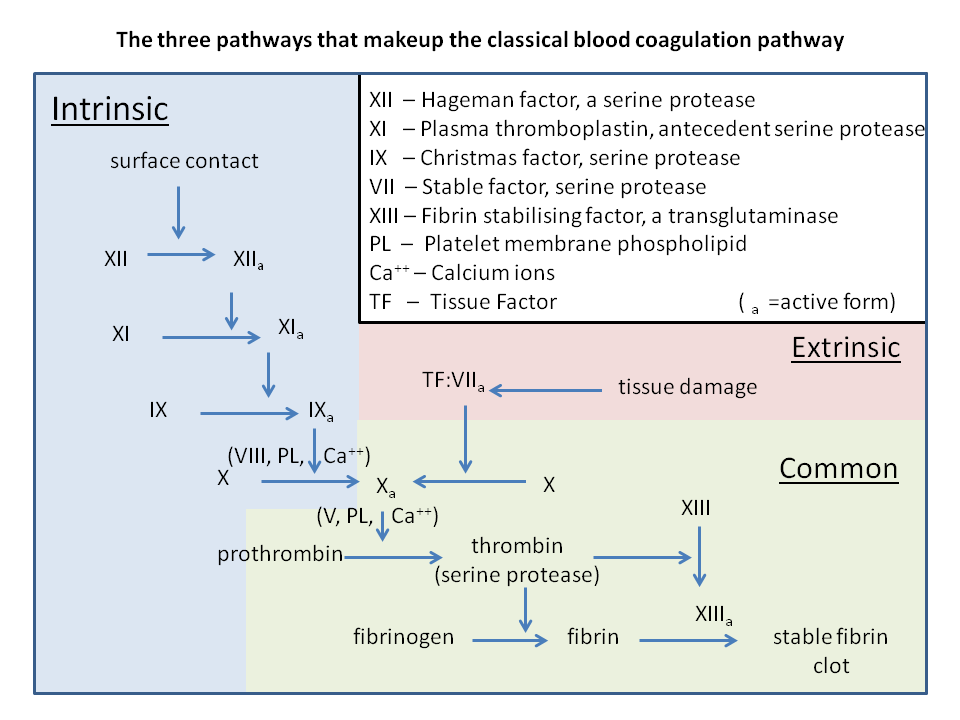
Laboratory assessment relies on mapping specific functional divisions within the enzymatic pathways to guide appropriate diagnostic testing:
- Intrinsic Pathway (Factors XII, XI, IX, VIII): Evaluated using the Activated Partial Thromboplastin Time (aPTT). This pathway is initiated when factor XII contacts a negatively charged surface.
- Extrinsic Pathway (Factor VII and Tissue Factor): Evaluated using the Prothrombin Time (PT) and International Normalized Ratio (INR). This pathway is triggered when trauma exposes tissue factor to the circulation.
- Common Pathway (Factors X, V, II [Prothrombin], I [Fibrinogen]): Point of convergence where both pathways merge to generate thrombin, which cleaves fibrinogen into insoluble fibrin bands. Deficiencies in the common pathway prolong both the PT and PTT.
2. Hereditary and Acquired Coagulation Defects
Distinguishing specific factor abnormalities relies heavily on inheritance patterns, clinical triggers, and exact laboratory cross-mixing trials:
| Condition | Molecular Defects & Pathophysiology | Lab Panel & 1:1 Mixing Study Response |
|---|---|---|
| Hemophilia A | • X-linked recessive deficiency of functional Factor VIII. • Disrupts the assembly of the intrinsic tenase complex, preventing downstream factor X activation. |
• Prolonged PTT; Normal PT and platelet count. • Corrects completely back to normal limits upon 1:1 mixing with normal pooled plasma. |
| Hemophilia B (Christmas Disease) |
• X-linked recessive deficiency of functional Factor IX. • Clinically identical to Hemophilia A in presentation, requiring specialized individual factor assays to distinguish. |
• Prolonged PTT; Normal PT and platelet count. • Corrects completely back to normal limits upon 1:1 mixing with normal pooled plasma. |
| Hemophilia C | • Autosomal recessive deficiency of Factor XI. • Found predominantly in individuals of Ashkenazi Jewish descent. Typically presents with mild, unprovoked bleeding or post-surgical hemorrhage. |
• Prolonged PTT; Normal PT. • Corrects completely back to normal limits upon 1:1 mixing with normal pooled plasma. |
| Coagulation Factor Inhibitors | • Acquired neutralizing IgG autoantibodies targeting a specific coagulation factor (most commonly anti-Factor VIII autoantibodies). • Can present clinically identical to severe hemophilia. |
• Prolonged PTT. • Fails to correct to normal limits upon 1:1 mixing because the autoantibodies neutralize the added factors. |
| Vitamin K Deficiency | • Impairs the gamma-glutamyl carboxylase enzyme, preventing post-translational modification of Factors II, VII, IX, X, and Proteins C and S. • Triggered by prolonged antibiotic use, malabsorption, or newborn status (due to a sterile gut). |
• Prolonged PT/INR (due to the short half-life of Factor VII). • Prolonged PTT follows in severe deficiency states. • Normal platelet count. |
3. Mixed Coagulation Profiles: Consumptive and Organ States
Several clinical scenarios cause profound secondary defects by combining structural organ failure with rapid element consumption:
- Disseminated Intravascular Coagulation (DIC):
Pathologic, systemic activation of the coagulation cascade, typically triggered by tissue factor release from sepsis (endotoxin), trauma, obstetric complications (amniotic fluid emboli), or acute leukemia (APL). This widespread microthrombi formation consumes platelets and coagulation factors, leading to simultaneous diffuse thrombosis and severe hemorrhage from IV lines and mucosal sites.• Lab Profile: Elevated PT/INR, elevated PTT, decreased platelet count, decreased fibrinogen, and markedly elevated D-dimer (indicating active fibrin degradation). Peripheral smear reveals prominent schistocytes. - Advanced Liver Disease:
The liver synthesizes nearly all circulating clotting factors and components of the fibrinolytic system. Advanced cirrhosis leads to decreased factor synthesis (except Factor VIII, which is produced by endothelial cells) and reduced thrombopoietin production, causing a combined quantitative deficiency.• Lab Profile: Prolonged PT/INR and aPTT, with concurrent thrombocytopenia due to hypersplenism from portal hypertension. Unlike DIC, D-dimer and fibrin degradation products remain near baseline or only mildly elevated.
You must be logged in to post a comment.