
Amyloidosis: High-Yield Pathology
Amyloidosis is a complex disease characterized by the extracellular deposition of abnormal, misfolded, insoluble protein fibrils. These fibrils aggregate into a cross-β-pleated sheet conformation, causing pressure atrophy and structural destruction of neighboring parenchymal cells.
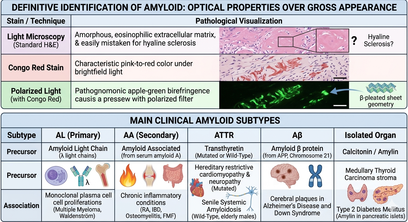
1. Staining & Diagnostic Hallmarks
The definitive identification of amyloid rests entirely on specific optical properties rather than gross appearance:
| Stain / Technique | Pathological Visualization |
|---|---|
| Light Microscopy | Appears as an amorphous, eosinophilic, acellular extracellular matrix under standard Hematoxylin & Eosin (H&E). Can easily be mistaken for hyaline sclerosis. |
| Congo Red Stain | Demonstrates a characteristic pink-to-red color under standard brightfield light microscopy. |
| Polarized Light | When Congo Red-stained tissue is split with polarized filters, it reveals a pathognomonic apple-green birefringence. This optical behavior is caused by the regular physical geometry of the β-pleated sheets. |
2. Main Clinical Subtypes
| Subtype | Precursor Protein | Classic Clinical Etiology & Associations |
|---|---|---|
| AL (Primary) |
Amyloid Light Chain (typically λ light chains) |
Caused by monoclonal plasma cell proliferations. Highly associated with Multiple Myeloma and Waldenström macroglobulinemia. |
| AA (Secondary) |
Amyloid Associated (cleaved from serum amyloid A) |
Driven by long-standing chronic inflammatory conditions. Classically seen in Rheumatoid Arthritis, Inflammatory Bowel Disease, Osteomyelitis, and Familial Mediterranean Fever (FMF). |
| ATTR | Transthyretin (Mutated or Wild-Type) |
• Mutated ATTR: Hereditary systemic amyloidosis causing restrictive cardiomyopathy and neuropathy. • Wild-Type ATTR: Senile Systemic Amyloidosis, predominantly accumulating in the hearts of elderly males. |
| Aβ | Amyloid β protein | Derived from the amyloid precursor protein (APP) on Chromosome 21. Forms the core of cerebral plaques in Alzheimer’s Disease and Down Syndrome. |
| Isolated Organ | Calcitonin / Amylin | • **Medullary Thyroid Carcinoma:** Tumor stroma shows amyloid from procalcitonin. • **Type 2 Diabetes Mellitus:** **Amylin (IAPP)** deposits damage pancreatic islet cells. |
3. Organ Manifestations & Morphology
- Kidneys: The most common and serious site of systemic involvement. Deposits settle initially within the glomeruli (mesangium and basement membranes), causing massive capillary permeability shifts. Clinically manifests as severe **Nephrotic Syndrome** progressing to chronic renal failure.
- Spleen: Produces two unique gross architectural patterns:
- Sago Spleen: Deposits are strictly confined to the splenic follicles, appearing as pale, translucent granules mimicking sago grains.
- Lardaceous Spleen: Deposits target the red pulp sinusoids, preserving the follicles but mapping into broad, map-like waxy zones.
- Heart: Classically causes a rigid, non-compliant ventricular wall leading to **Restrictive Cardiomyopathy** and conduction system arrhythmias. On echocardiography, it displays a characteristic “speckled” or “granular” myocardial pattern.
- Gastrointestinal: Macroglossia (massive enlargement of the tongue) is a highly specific physical sign pointing toward AL amyloidosis. Malabsorption can occur if fibrils infiltrate the small intestinal villi.
4. High-Yield Exam Pearls
- Screening Site: The preferred, least invasive clinical site for screening systemic amyloidosis is an **Abdominal Fat Pad Aspirate/Biopsy** or a rectal biopsy, both showing high diagnostic sensitivity when stained with Congo Red.
- $\beta_2$-Microglobulin Amyloidosis: Occurs in patients on long-term **hemodialysis**. The protein cannot pass through standard dialysis membranes, leading to systemic accumulation with a unique tropism for joints, presenting as **Carpal Tunnel Syndrome**.
You must be logged in to post a comment.