M04.01.007 Ultrastructure of Blood Vessels
Blood vessels are vital components of the circulatory system, responsible for transporting blood throughout the body. They are categorized into three main types: arteries, veins, and capillaries. Each type has distinct anatomical and histological features, and their clinical relevance lies in their role in various diseases and medical interventions. Let’s explore each aspect in detail:
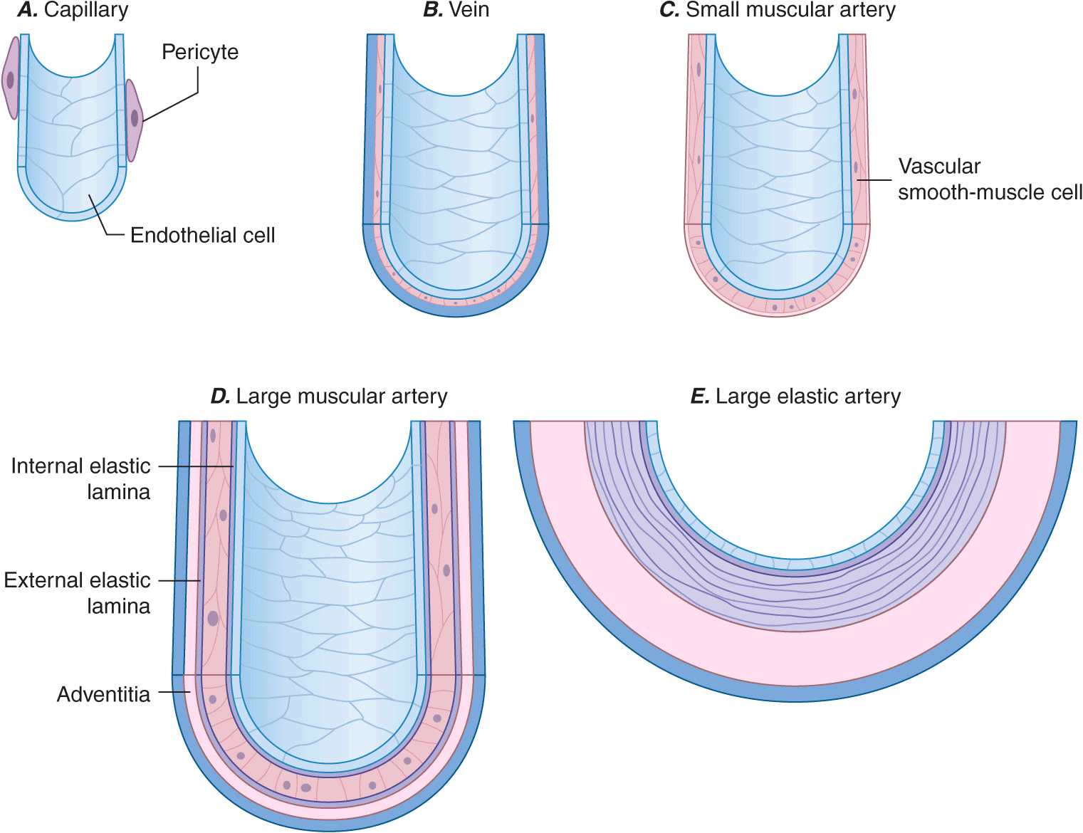
Anatomy of Blood Vessels:
Arteries: Arteries carry oxygenated blood away from the heart to the various tissues and organs of the body. They have thick, elastic walls composed of three layers: the innermost tunica intima, the middle tunica media (with smooth muscle cells and elastic fibers), and the outer tunica adventitia (composed of connective tissue).
Veins: Veins transport deoxygenated blood back to the heart. They have thinner walls compared to arteries and contain valves that prevent the backflow of blood. Veins consist of the same three layers as arteries, but their tunica media is less developed, and the tunica adventitia is more prominent.
Capillaries: Capillaries are the smallest and most numerous blood vessels. They connect arteries and veins, enabling the exchange of oxygen, nutrients, waste products, and hormones with the surrounding tissues. Capillary walls are composed of a single layer of endothelial cells, allowing for easy diffusion of substances between the blood and tissues.
Histology of Blood Vessels:
Arteries: The tunica intima of arteries consists of a thin layer of endothelial cells surrounded by a subendothelial layer of connective tissue. The tunica media contains smooth muscle cells, elastic fibers, and collagen. The tunica adventitia is composed of connective tissue with collagen and elastic fibers.
Veins: Veins have a similar structure to arteries but with thinner walls. The tunica intima is also composed of endothelial cells and a subendothelial layer. The tunica media contains smooth muscle cells, although it is less developed compared to arteries. The tunica adventitia is thicker and contains collagen and elastic fibers.
Capillaries: Capillaries consist of a single layer of endothelial cells, which are thin and permit the exchange of substances between blood and tissues. Some capillaries are fenestrated (contain pores) or have gaps between endothelial cells, facilitating the passage of larger molecules.

Clinical Relevance of Blood Vessels:
Atherosclerosis: Atherosclerosis is a common condition characterized by the buildup of plaque inside arterial walls. This can lead to narrowing or blockage of arteries, reducing blood flow and causing various cardiovascular diseases, such as coronary artery disease, stroke, or peripheral artery disease.
Hypertension: High blood pressure can damage the walls of blood vessels, particularly small arterioles, and capillaries. This can contribute to the development of conditions such as retinopathy, nephropathy, and peripheral vascular disease.
Deep Vein Thrombosis (DVT): DVT occurs when a blood clot forms in a vein, often in the lower limbs. It can lead to complications such as pulmonary embolism if the clot dislodges and travels to the lungs. Understanding the anatomy and histology of veins is important in diagnosing and managing DVT.
Angiography: Angiography is a medical imaging technique used to visualize blood vessels. It involves the injection of contrast dye into the bloodstream, which highlights the blood vessels and helps detect abnormalities such as blockages or aneurysms.
Vascular Surgery: Knowledge of blood vessel anatomy and histology is crucial in vascular surgery, including procedures such as bypass grafting
You must be logged in to post a comment.