Learning Objectives
Identify the three primary layers of the skin and master the five distinct sub-layers of the epidermis. Understand the structural progression of keratinocytes from the regenerative stratum basale to the protective stratum corneum.
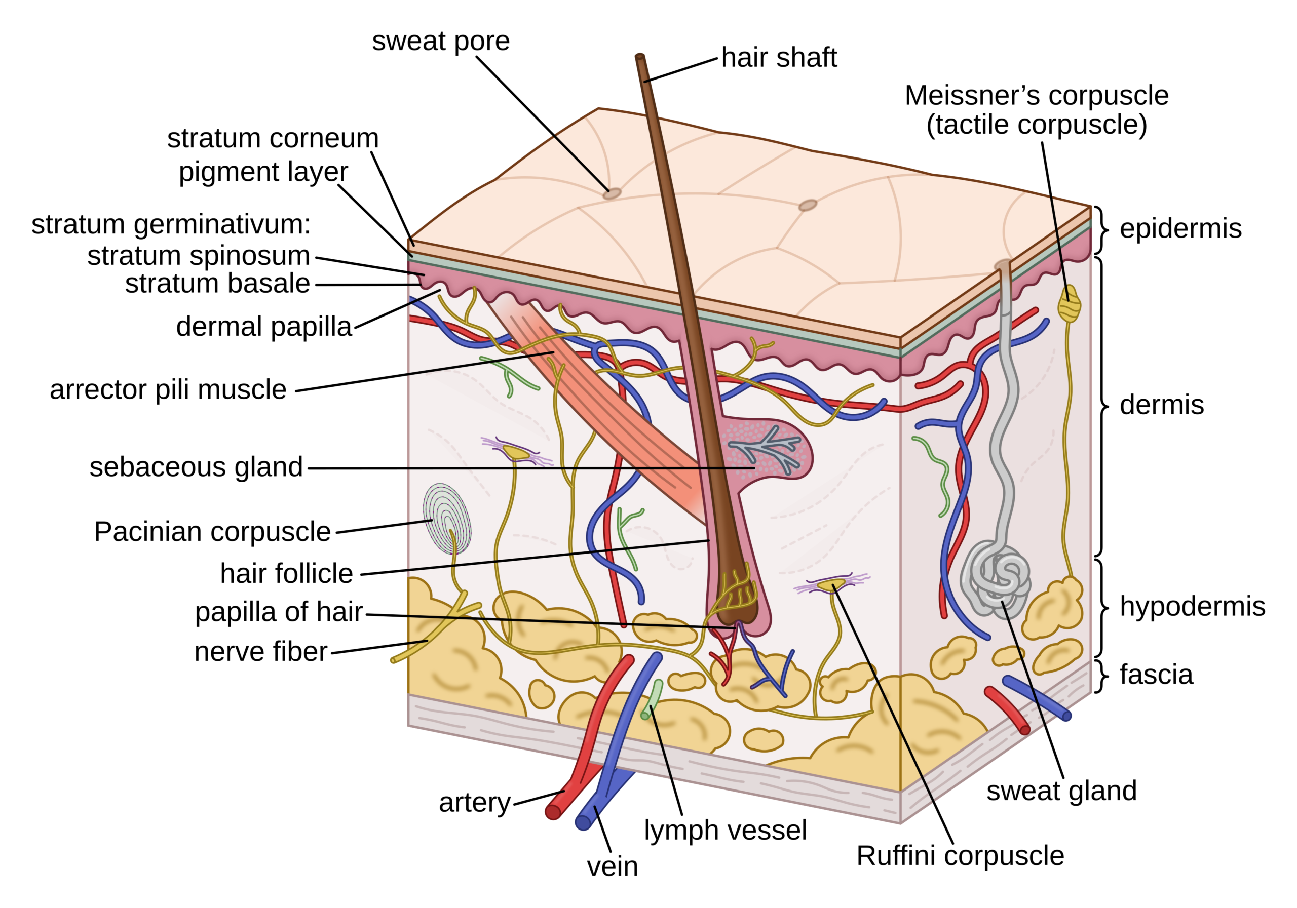
1. Primary Skin Layers
The skin (integumentary system) is composed of three functional layers that vary in thickness and composition across the body.
- Epidermis: The outermost, avascular layer consisting mainly of keratinocytes.
- Dermis: The middle layer containing connective tissue, blood vessels, nerves, hair follicles, and sweat glands.
- Subcutaneous Fat (Hypodermis/Subcutis): The deepest layer, composed of adipose and loose connective tissue for insulation and shock absorption.

2. The Epidermal Layers (Stratification)
The epidermis is further divided into five strata. Cells migrate upward from the basement membrane, maturing and eventually desquamating (shedding).
| Layer | Key Characteristics | High-Yield Note |
|---|---|---|
| Stratum Corneum | Top layer: dead, keratin-filled cells (corneocytes). | Provides the primary barrier function. |
| Stratum Lucidum | Clear, thin layer. | Found only in thick skin (palms and soles). |
| Stratum Granulosum | Cells contain keratohyalin granules. | Where keratinocytes begin to lose their nuclei. |
| Stratum Spinosum | “Spiny” layer due to desmosomes holding cells together. | Thickest layer in most skin areas. |
| Stratum Basale | Deepest, single layer of mitotically active cells. | Contains stem cells and melanocytes. |
Activity: Microscopic Identification of Skin Strata
High-Yield Mnemonic:To remember the layers from Superficial to Deep (Top to Bottom):
- Come: Corneum
- Let’s: Lucidum
- Get: Granulosum
- Sun: Spinosum
- Burned: Basale
You must be logged in to post a comment.