Learning Objectives
- Identify the vascular supply to the femoral head and the primary artery involved in necrosis.
- Recall the high-yield mnemonic (CASTS Bend LEGS) for the causes of avascular necrosis.
- Explain the concept of a watershed area and why it is prone to infarction.
- Recognize the clinical presentation and radiographic features of bone infarction.
1. Pathophysiology: Bone Infarction
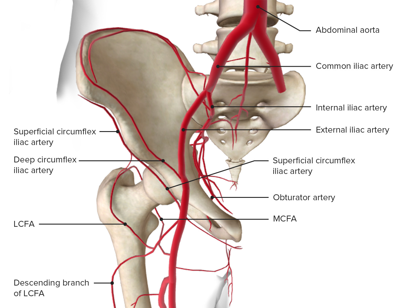
Avascular necrosis (AVN), or osteonecrosis, is the death of bone tissue due to a lack of blood supply. It most commonly occurs in the femoral head because of its precarious “watershed” vascularity.
- The Vulnerable Vessel: The Medial Circumflex Femoral Artery provides the majority of the blood supply to the femoral head. Insufficiency or damage to this artery is the most common cause of AVN.
- Watershed Area: The subchondral bone of the femoral head is a “terminal” zone with limited collateral circulation, making it the first area to die during ischemia.
2. Causes of Avascular Necrosis
The etiology of AVN is varied, ranging from direct trauma to systemic diseases that impair microcirculation.
| Letter | Cause | High-Yield Mechanism |
|---|---|---|
| C | Corticosteroids | Chronic glucoCorticoid use is the most common non-traumatic cause. |
| A | Alcohol | Chronic overuse leads to fat emboli or altered lipid metabolism. |
| S | Sickle Cell Disease | Vaso-occlusive crises in the bone marrow. |
| T | Trauma | Femoral neck fractures can disrupt the medial circumflex artery. |
| S | SLE | Systemic Lupus Erythematosus (often combined with steroid use). |
| Bend | The Bends | Caisson disease; nitrogen bubbles embolize in the bone. |
| LEGS | Legg-Calvé-Perthes / SCFE | Pediatric hip disorders involving femoral head ischemia. |
3. Clinical Features & Diagnosis
AVN presents as progressive, deep-seated pain that is worse with weight-bearing and movement.
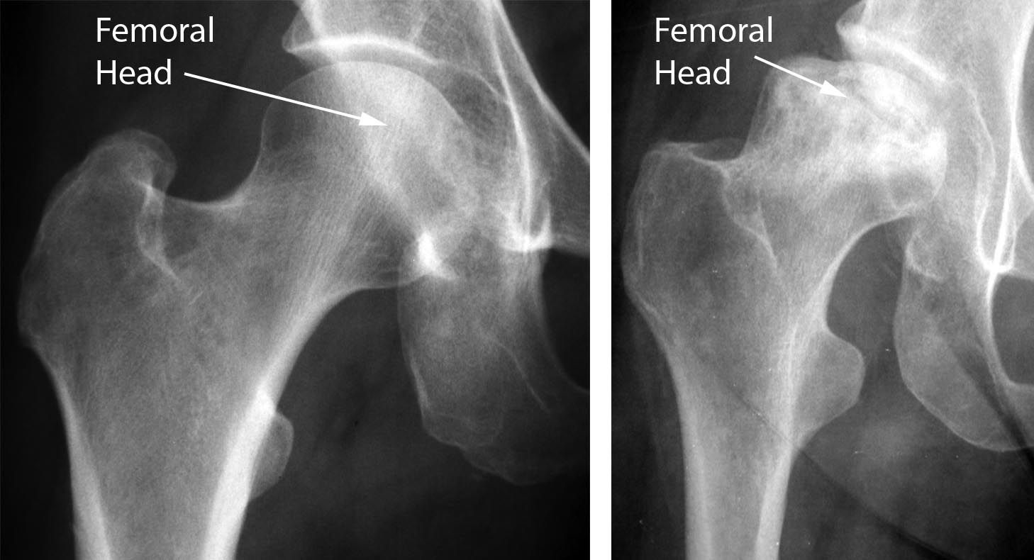
- Imaging:
- MRI: The most sensitive test for early-stage AVN.
- X-ray: May show “creeping substitution” or the classic Crescent Sign (subchondral collapse/fracture).
- Physical Exam: Marked limitation of internal rotation and abduction of the hip.
Clinical Notes & Step 1 Pearls:
- Gaucher Disease: Don’t forget that AVN is a classic complication of Gaucher disease due to glucocerebroside accumulation in the bone marrow.
- Scaphoid Bone: After the femoral head, the scaphoid bone is the second most common site for AVN following a fracture (distal-to-proximal blood supply).
- Outcome: If untreated, AVN leads to progressive joint destruction and secondary osteoarthritis, eventually requiring a total hip arthroplasty.
Activity: AVN Etiology Challenge
Quick Mnemonic:
CASTS Bend LEGS:
Corticosteroids, Alcohol, Sickle cell, Trauma, SLE, the Bends, Legg-Calvé-Perthes, Gaucher, SCFE.
You must be logged in to post a comment.