Learning Objectives
- Identify the most common site of clavicle fractures and the underlying anatomy.
- Explain the mechanical forces (muscles) that cause bone displacement after a fracture.
- Recognize the clinical presentation of clavicular trauma in pediatric and neonatal populations.
1. Clavicle Fracture Anatomy
The clavicle is the first bone to ossify in the fetus and the most commonly fractured bone during birth and childhood.
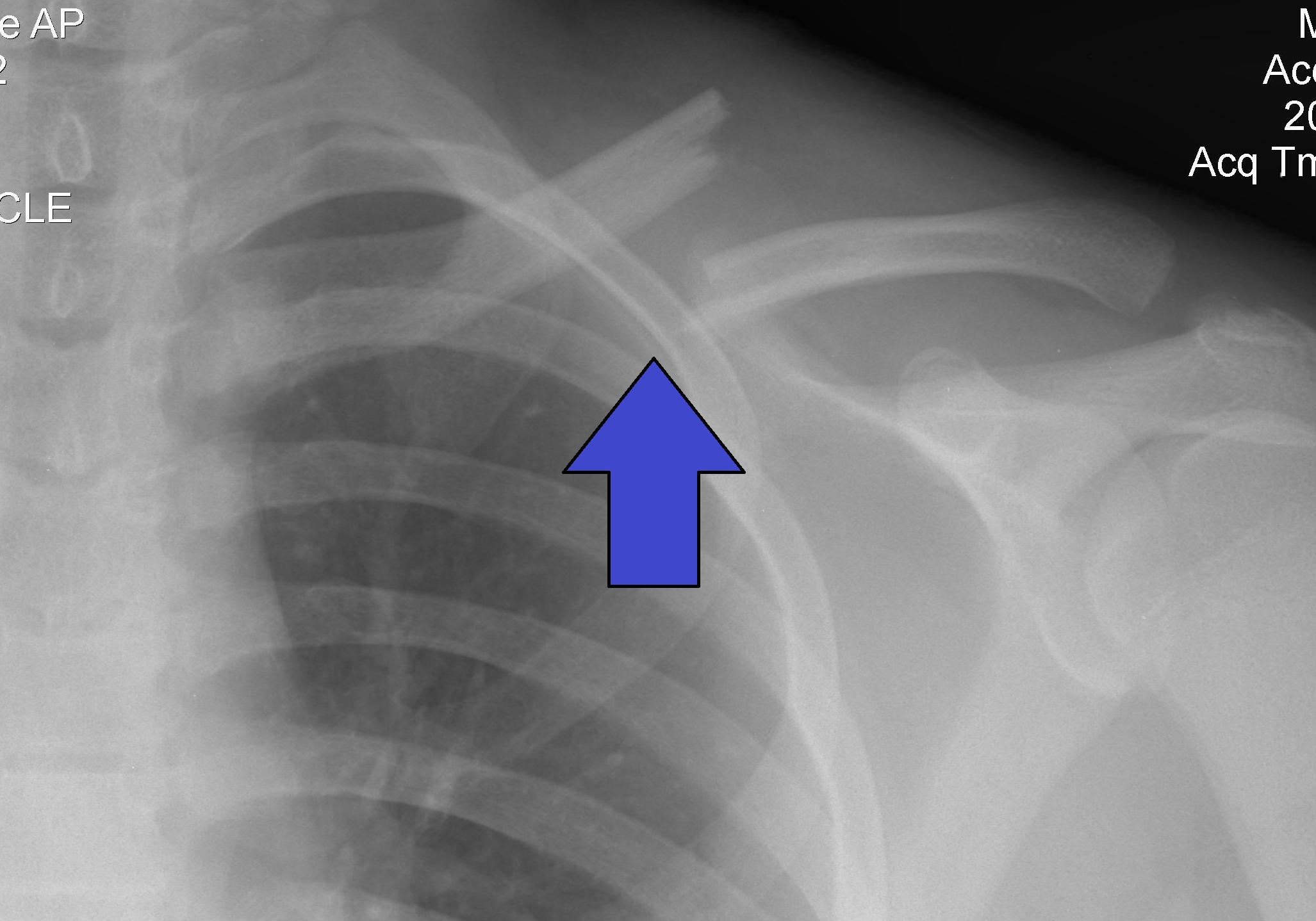
- Weakest Point: The junction of the middle and lateral thirds. This is where the curvature of the bone changes and where most fractures occur.
- Mechanism: A fall on an outstretched hand (FOOSH), direct trauma to the shoulder, or birth trauma (macrosomia/shoulder dystocia).
2. Clinical Presentation & Muscle Pull
The characteristic “deformity” of a clavicle fracture is caused by the antagonistic pull of various muscles attached to the bone fragments.
| Fragment | Displacement Direction | Responsible Muscle |
|---|---|---|
| Medial Fragment | Superior (Upward) | Sternocleidomastoid (SCM) |
| Lateral Fragment | Inferior (Downward) | Weight of the arm (Gravity) |
| Lateral Fragment | Medial (Inward) | Pectoralis major (Adduction) |
- Physical Exam: The patient will present with a “shoulder drop” and will often support the affected arm with the opposite hand. The clavicle may appear shortened due to the medial overlap of the lateral fragment.
Clinical Notes & Step 1 Pearls:
- Vascular Risk: The Subclavian artery and the Brachial plexus run directly posterior to the middle third of the clavicle. A severely displaced fracture or a posterior fragment can cause neurovascular injury (check distal pulses!).
- Neonatal Presentation: A newborn with a clavicle fracture may present with pseudoparalysis (refusal to move the arm due to pain) and a localized “lump” (callus formation) a few weeks later.
- Ossification: Remember that the clavicle undergoes intramembranous ossification, despite being a long bone.
Activity: Clavicle Displacement Challenge
Quick Mnemonics:
SCM: Pulls the Stubborn Central (Medial) part Moving up.
Pec Major: Pulls the Peripheral (Lateral) part Medially.
You must be logged in to post a comment.