Learning Objectives
- Differentiate between Low Ankle Sprains and High Ankle Sprains.
- Identify the Anterior Talofibular Ligament (ATFL) as the most commonly injured structure.
- Explain the mechanism of injury (inversion vs. rotation) for different ligament tears.
- Map the bones of the foot, including tarsals, metatarsals, and phalanges.
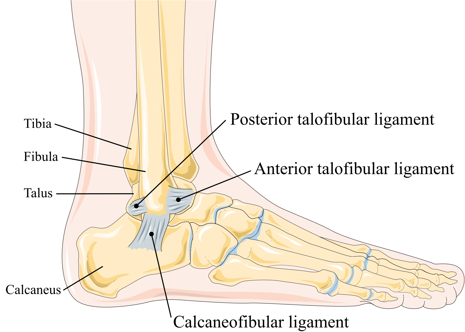
1. Low Ankle Sprains (Lateral Ligaments)
Low ankle sprains involve the lateral ligament complex. These are almost always caused by overinversion and supination of the foot.
- Anterior Talofibular Ligament (ATFL): The most common ankle sprain overall. It is the first ligament to tear during an inversion injury.
- Calcaneofibular Ligament (CFL): The second most common; usually injured if the inversion force continues after the ATFL has torn.
- Posterior Talofibular Ligament (PTFL): The strongest of the lateral ligaments; rarely injured except in complete dislocations.
2. High Ankle Sprains (Syndesmotic)
High ankle sprains involve the tibiofibular syndesmosis, which holds the tibia and fibula together above the talus.
- Anterior Inferior Tibiofibular Ligament (AITFL): The most common high ankle sprain.
- Mechanism: Usually caused by external rotation of the foot or extreme dorsiflexion, which spreads the tibia and fibula apart.
- Clinical: These take significantly longer to heal than low ankle sprains because they are weight-bearing structures.
3. Anatomy of the Foot Bones
The foot is divided into the tarsals (ankle/midfoot), metatarsals, and phalanges.
- Tarsals: Talus (articulates with tibia/fibula), Calcaneus (heel), Navicular, Cuboid, and the three Cuneiforms (Medial, Intermediate, Lateral).
- Metatarsals: Numbered 1–5 starting from the big toe (hallux).
- Phalanges: Proximal, middle, and distal (the big toe only has proximal and distal).
Activity:
Clinical Notes & Corrections:
- Anterior Drawer Test (Ankle): Used specifically to assess the integrity of the ATFL. A positive test shows the talus sliding forward under the tibia.
- Medial Ligaments: The Deltoid ligament on the medial side is very strong. Medial sprains are rare; instead, the force often results in an avulsion fracture of the medial malleolus.
- Ottawa Ankle Rules: Used in the ER to determine if an X-ray is needed based on bone tenderness (malleoli, navicular, or 5th metatarsal) and the ability to bear weight.
Activity: Ankle Ligament Identification
Memory Hooks:
ATFL: Always Tears First Laterally.
High Tide: High ankle sprains = Tibiofibular ligaments (Tib sounds like Tide).
Inversion: Most sprains go In (Inversion), hitting the Outside (Lateral) ligaments.
Activity:
You must be logged in to post a comment.