Learning Objectives
Master the mechanisms of Free Radical Injury and their role in cellular pathology. Identify the pathways of radical initiation—including the Fenton reaction and drug metabolism—and recognize the essential antioxidant defense systems used to neutralize oxidative stress.
1. Mechanisms of Free Radical Damage
Free radicals are highly reactive molecules with an unpaired electron in their outer orbit. They trigger a chain reaction of cellular destruction through three primary pathways:
| Damage Target | Consequence |
|---|---|
| Lipid Peroxidation | Oxidation of polyunsaturated fats → Membrane damage (plasma and organelle). |
| Protein Modification | Oxidation of amino acid side chains → Protein misfolding and enzyme inactivation. |
| DNA Breakage | Reaction with thymine → Single-strand breaks; linked to aging and malignant transformation. |
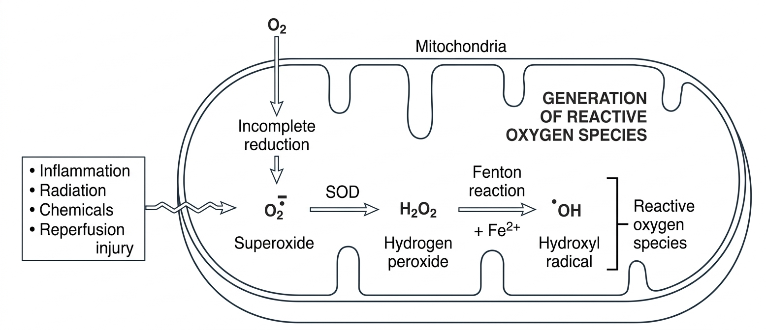
2. Initiation and Clinical Examples
Radicals are generated during normal metabolism but can reach toxic levels via exogenous triggers or metabolic failure.
| Source | Process / Mechanism | Clinical Context |
|---|---|---|
| Transition Metals | Fenton Reaction:  or $Cu^{2+}$ generate or $Cu^{2+}$ generate  . . |
Hemochromatosis (Iron) or Wilson Disease (Copper). |
| Chemical Toxicity | P-450 conversion of  to $CCl_3^\bullet$. to $CCl_3^\bullet$. |
Fatty liver (due to ↓ apolipoprotein synthesis). |
| Drug Overdose | Metabolism into toxic intermediates. | Acetaminophen hepatotoxicity. |
| Oxygen Toxicity | High $O_2$ concentrations in neonates. | Retinopathy of prematurity; Bronchopulmonary dysplasia. |
Activity:
3. Elimination and Antioxidant Defense
The body uses enzymatic and non-enzymatic scavengers to prevent radical-mediated injury.
- Enzymes: Superoxide Dismutase (
), Catalase (
), and Glutathione Peroxidase.
- Vitamins: Vitamins A, C, and E act as direct antioxidants.
- Carrier Proteins: Transferrin and Ceruloplasmin sequester metals to prevent the Fenton reaction.
Activity
High-Yield Mnemonics & Tips:
- CCl4 Fatty Liver: Remember that $CCl_4$ injury causes ribosomal detachment. No ribosomes = no apolipoproteins. Without apolipoproteins, fat can’t leave the liver, leading to steatosis.
- Reperfusion Wave: Returning blood to an ischemic area brings oxygen and neutrophils, which combine to create a “burst” of free radicals, often worsening the initial damage.
- The Fenton Reaction: Just remember “Iron (Fe) makes Free radicals.” This is the core mechanism behind the organ damage seen in hemochromatosis.
You must be logged in to post a comment.