Learning Objectives
Master the structural and functional organization of the Lymph Node. Identify the specific histological zones—the Follicle, Paracortex, and Medulla—and correlate their structure with the circulation and activation of B and T cells for the USMLE Step 1.
1. General Lymph Node Structure
A lymph node is an encapsulated secondary (
| Feature | Description |
|---|---|
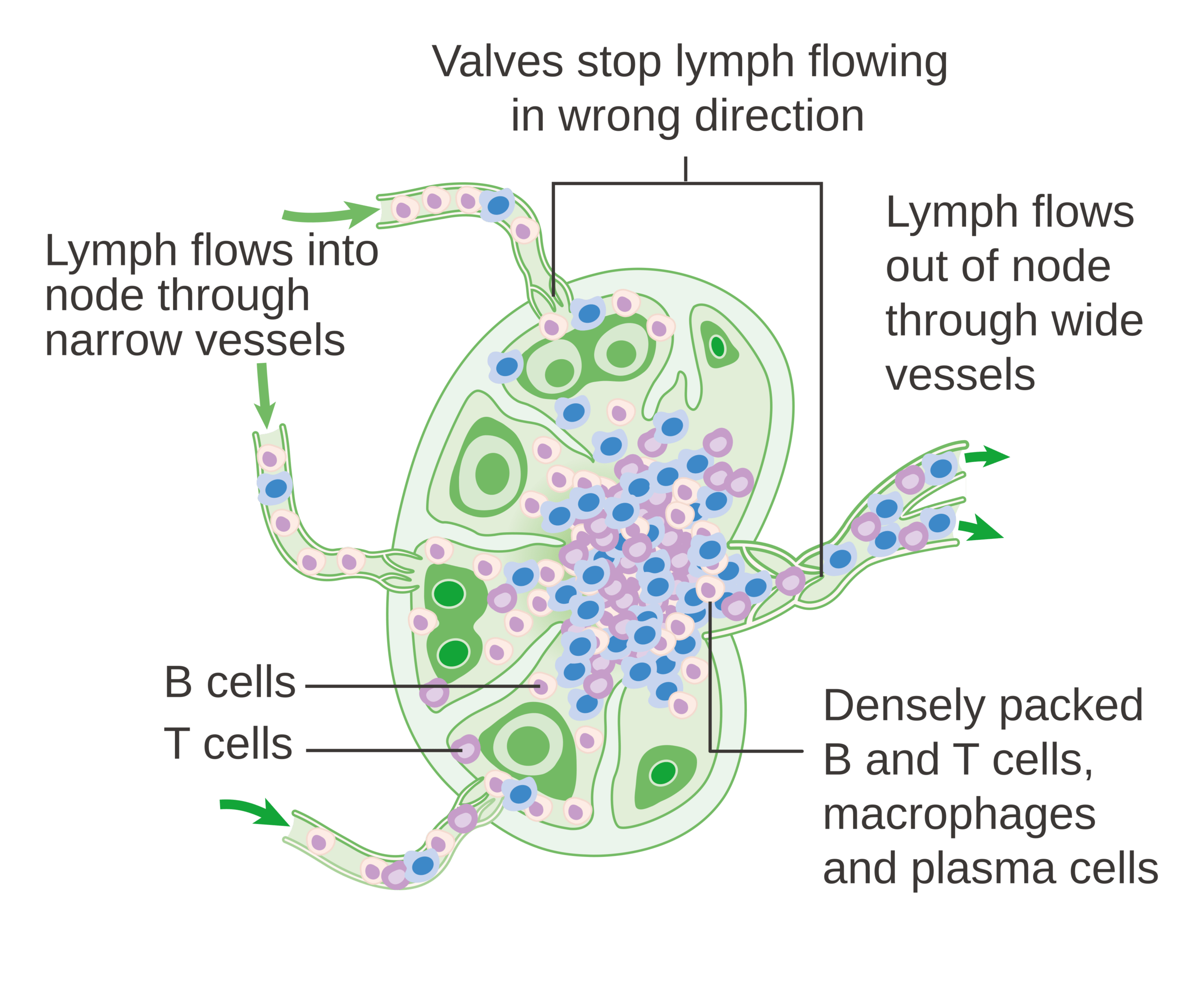
| Vascularity | Many afferent vessels (entry);  or more efferent vessels (exit). or more efferent vessels (exit). |
| Architecture | Encapsulated, with trabeculae (structural partitions) providing internal support. |
| Functions | Nonspecific filtration by macrophages, circulation of lymphocytes, and activation of the immune response. |
2. The Cortex and Follicles (B-Cell Zone)
The outer cortex is primarily composed of follicles, which are the sites of B-cell localization and proliferation.
| Follicle Type | Characteristics |
|---|---|
 Follicles Follicles |
Dense and quiescent (inactive). |
| Follicles |
Active; characterized by pale central germinal centers where B cells proliferate and undergo isotype switching. |
3. The Paracortex (T-Cell Zone)
The paracortex is the region between the outer cortex (follicles) and the inner medulla. It is the primary site for T-cell interaction.
| Anatomy/Clinical | Significance |
|---|---|
| High Endothelial Venules (HEV) | The specialized entry point where T and B cells exit the blood and enter the lymph node. |
| DiGeorge Syndrome | The paracortex is underdeveloped due to a lack of mature T cells. |
| Viral Infection (EBV) | Triggers an extreme cellular immune response, causing paracortical hyperplasia and lymphadenopathy. |
4. The Medulla
The innermost part of the lymph node consists of cords and sinuses that facilitate filtration and exit.
| Region | Contents |
|---|---|
| Medullary Cords | Closely packed lymphocytes and plasma cells. |
| Medullary Sinuses | Contain reticular cells and macrophages. These communicate directly with efferent lymphatics. |
Activity
High-Yield Clinical Pearls:
- Paracortex vs. Cortex: Remember that the paracortex is for T cells, and the follicles in the cortex are for B cells. If T cells are low (HIV, DiGeorge), look at the paracortex; if antibodies are low, look at the follicles.
- Lymph Flow: Afferent lymphatic
Subcapsular sinus
- Viral Hyperplasia: Unlike bacterial infections, which often cause follicular hyperplasia (B cells), viral infections such as Mononucleosis often cause paracortical hyperplasia (T cells).
You must be logged in to post a comment.