Learning Objectives
- Compare the kinetic properties (
and
) of Hexokinase and Glucokinase.
- Identify the tissue distribution and metabolic purpose of each enzyme.
- Understand how insulin and feedback mechanisms regulate glucose trapping.
1. Glucose Trapping Mechanism
The phosphorylation of glucose to Glucose-6-Phosphate (G6P) is the first step of glycolysis. This step “traps” glucose inside the cell, as G6P cannot cross the plasma membrane.
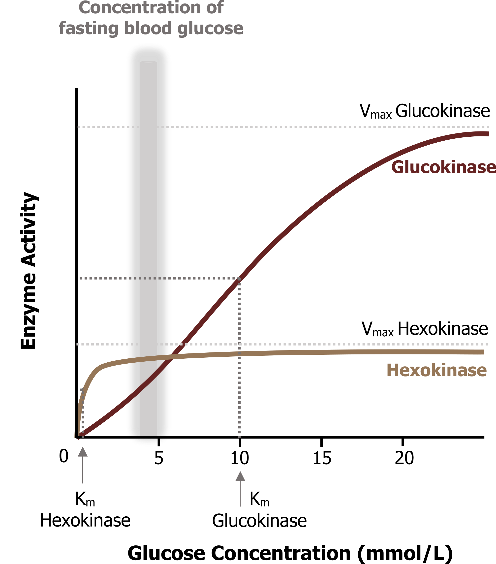
| Feature | Hexokinase | Glucokinase (Hexokinase IV) |
|---|---|---|
| Location | Most tissues (except liver/β cells) | Liver and β cells of the pancreas |
| Affinity ($Latex K_m$) | Lower $Latex K_m$ (Higher affinity) | Higher $Latex K_m$ (Lower affinity) |
| Capacity ($LatexV_{max}$) | Lower $Latex V_{max}$ | Higher $Latex V_{max}$ |
| Insulin Induction | No | Yes |
| Feedback Inhibition | Glucose-6-phosphate | Fructose-6-phosphate |
Biochemical Correlation: and Physiological Roles
Hexokinase’s low
Clinical Correlate: Glucokinase Deficiency
Deficiency in glucokinase raises the threshold for glucose-stimulated insulin release. This is a primary cause of Maturity-Onset Diabetes of the Young (MODY) and Gestational Diabetes, as β cells require higher glucose concentrations to trigger insulin secretion.
Activity
You must be logged in to post a comment.