Learning Objectives
By the end of this session, the learner will be able to identify major cortical landmarks and layers, localize language functions in the dominant hemisphere, interpret homunculus organization, understand cerebral blood supply and vascular territories, and correlate lesions with clinical stroke syndromes and cortical deficits.
1. General Features: Gross Anatomy
The cerebral cortex is highly folded into gyri (ridges) and sulci (grooves), increasing functional surface area.
| Landmark | Anatomical Role | Clinical Importance |
|---|---|---|
| Lateral Fissure (Sylvian) | Separates the temporal from the frontal/parietal lobes | MCA runs here → stroke affects language |
| Central Sulcus | Separates frontal (motor) and parietal (sensory) | Key landmark for motor vs sensory deficits |
| Cingulate Sulcus | Separates the cortex from the limbic lobe | Important in emotion/behavior circuits |
| Parieto-occipital Sulcus | Separates parietal and occipital lobes | Visual vs sensory cortex boundary |
| Calcarine Sulcus | Primary visual cortex location | Lesion → homonymous hemianopia |
2. Cortical Histology & Layers
The cortex is functionally organized into layers that reflect input vs output roles.
| Layer | Function | Clinical Relevance |
|---|---|---|
| I–III | Association & integration | Cortical processing |
| IV | Receives thalamic input | Primary sensory cortex prominent |
| V | Motor output (corticospinal) | Damage → paralysis (Betz cells) |
| VI | Projects back to the thalamus | Feedback regulation |
Key Distinction:
- Neocortex: 6 layers (90%)
- Allocortex: 3 layers (hippocampus, olfactory)
3. Language & Dominant Hemisphere
Dominance: Left hemisphere in most individuals
[Image of the brain showing Broca’s and Wernicke’s areas]
| Condition | Location | Key Clinical Features |
|---|---|---|
| Broca Aphasia | Frontal (44,45) | Non-fluent speech, intact comprehension, frustrated |
| Wernicke Aphasia | Temporal (22) | Fluent but meaningless, poor comprehension, unaware |
| Conduction Aphasia | Arcuate fasciculus | Cannot repeat, fluent, aware |
| Gerstmann Syndrome | Angular gyrus | Agraphia, acalculia, finger agnosia, R-L confusion |
4. Frontal Lobe Functions
| Functional Region | Physiological Role | Lesion Presentation |
|---|---|---|
| Primary Motor (Area 4) | Executes contralateral voluntary movement | Contralateral spastic weakness |
| Premotor (Area 6) | Planning/sequencing of complex movements | Apraxia |
| Frontal Eye Field (8) | Contralateral horizontal saccades | Eyes deviate toward the lesion |
| Prefrontal Cortex | Executive function, judgment, and social behavior | Apathy, disinhibition, poor judgment |
5. Parietal Lobe Functions
| Functional Area | Normal Function | Lesion Presentation |
|---|---|---|
| Primary Sensory Cortex | Touch, pain, vibration, proprioception | Contralateral sensory loss |
| Association Cortex (5,7) | Sensory integration & spatial mapping | Astereognosis and Apraxia |
| Nondominant Parietal | Spatial awareness of the contralateral side | Hemispatial neglect syndrome |
6. Occipital & Temporal Lobes
| Lobe | Functional Region | Clinical Deficit |
|---|---|---|
| Occipital | Primary Visual Cortex (Area 17) | Contralateral hemianopia with macular sparing |
| Temporal | Wernicke’s Area (Superior Gyrus) | Receptive aphasia (fluent nonsense) |
| Temporal | Meyer’s Loop (Optic Radiation) | Contralateral superior quadrantanopia (“pie in sky”) |
7. Homunculus Organization
| Surface Aspect | Body Representation | Vascular Supply (Rule) |
|---|---|---|
| Lateral Convexity | Face and Upper Limb (Arm/Hand) | MCA → Face/arm deficits |
| Medial Surface | Lower Limb (Leg/Foot) and Perineum | ACA → Leg deficits |
8. Blood Supply: Circle of Willis
| System | Key Components | High-Yield Clinical Connection |
|---|---|---|
| Internal Carotid | ACA, MCA, ACoA | Anterior circulation; supplies 80% of the brain |
| Vertebrobasilar | Vertebral, Basilar, PCA | Posterior circulation; supplies the brainstem/occipital |
| Communicating | Ant. & Post. Communicating Arteries | PCoA Aneurysm → CN III palsy |
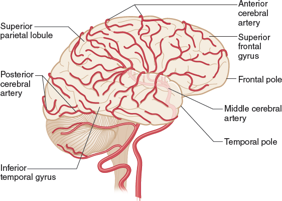
9. Major Arterial Syndromes
| Artery | Territory | Deficit |
|---|---|---|
| MCA | Lateral cortex | Face/arm paralysis, aphasia (L), neglect (R) |
| ACA | Medial cortex | Leg weakness, incontinence |
| PCA | Occipital | Hemianopia, macular sparing |
10. Hemorrhage Types
| Type | Cause | Clue |
|---|---|---|
| Epidural | MMA rupture | Lucid interval |
| Subdural | Bridging veins | Slow bleed |
| Subarachnoid | Berry aneurysm | Worst headache |
| Intracerebral | Hypertension | Deep bleed |
Check Your Knowledge:
Critical Concepts
- Left hemisphere = language
- Right parietal lesion = neglect
- MCA = most common stroke
- Internal capsule lesions = dense deficits
Clinical Pearls:
- Broca vs Wernicke:
Broca = Broken speech
Wernicke = Fluent nonsense - MCA Rule:
Face/arm > leg - ACA Rule:
Leg > arm - PCA Rule:
Vision loss with macular sparing
You must be logged in to post a comment.